Total Shoulder Replacement or Reverse Shoulder Replacement
May 21, 2018
Shoulder arthroplasty, often called a total shoulder replacement, is a well-established surgery for treating severe pain and stiffness. In fact, approximately 53,000 people in the U.S. have shoulder replacement surgery each year in order to regain comfort and function, according to the Agency for Healthcare Research and Quality. And while the majority of these procedures would be considered standard replacements, patients may be surprised to know that a “reverse” replacement is also an option. In some situations, a reverse shoulder replacement may benefit a patient more than a standard procedure.
At Beacon Orthopaedics and Sports Medicine, our shoulder specialists have performed numerous total shoulder replacements and reverse shoulder replacements. If you are experiencing shoulder pain and stiffness that limits your activities during the day and keeps you awake at night, you should talk to a specialist about your options. Here is information about total shoulder replacements and reverse shoulder replacements to help guide your discussion.
Q: What is a total shoulder replacement?
In a healthy shoulder, the head of the upper arm bone (humerus) fits into a shallow socket in the scapula. In shoulder replacement surgery the damaged portions of the shoulder are removed and replaced with a smooth metal ball attached to a stem. This artificial component, called a prothesis, will either replace the “ball” of the joint or both the “ball” and “socket.”
Q: What is a reverse shoulder replacement?
 In reverse shoulder replacement surgery, the damaged portions of the shoulder are also replaced with a prosthesis made from metal and plastic. However, unlike a standard replacement, the artificial component is placed on the socket side of the joint. The socket is then placed on the arm side where it is supported by a metal stem. Thus, the prosthesis is reverse of where its natural counterparts would be located within the body.
Reverse shoulder replacement works best for patients with cuff tear arthropathy or a detached rotator cuff. The reverse prosthesis allows the patient to use their deltoid muscles instead of their deteriorated or detached rotator cuff tendons in order to lift their arm. It is also effective in people with complex fractures or people who have extensive bone loss.
Q: What are the benefits of shoulder replacement surgery?
 First and foremost, the purpose of shoulder surgery is to relieve chronic pain and restore mobility so patients can resume normal living. Additionally, an orthopedic surgeon may recommend a shoulder replacement in order to treat the following conditions:
- Osteoarthritis
- Rheumatoid Arthritis
- Post-traumatic arthritis
- Rotator Cuff Tear Arthropathy
- Avascular Necrosis
- Severe Fractures
- Bone Tumors
In recent years, advancements in prosthetic designs have allowed patients to resume active lifestyles following surgery. Patients can often return to baseball, golf, swimming, or many of the other activities they enjoy.
Q: How successful is shoulder replacement surgery?
Both total shoulder replacement and reverse shoulder replacement have been shown to be highly effective. As with all forms of surgery, however, the effectiveness of the procedure depends on the overall health of the patient, their participation in physical therapy, and the expertise of the orthopaedic surgeon. Patients can improve their outcomes by choosing an experienced orthopaedic surgeon and adhering to their treatment plan.
Q: What are the potential risks of shoulder surgery?
Shoulder replacements are considered as safe as hip and knee replacement surgeries. Of course, as with any joint replacement surgery, there are associated risks. The most common complication is the dislocation of the prosthesis, with dislocations being more likely to occur among reverse shoulder replacements. Fortunately, a dislocation can be managed by placing the arm back into the proper location and keeping the arm immobile for a period of time.
Shoulder replacement surgery also carries risks associated with other forms of surgery, including:
- Infection
- Fracture
- Nerve or blood vessel damage
- Joint stiffness or instability
- Loosening of the prothesis
- Dislocation
Although rare, other complications may occur. An experienced orthopaedic surgeon can talk to you about these complications and discuss your individual level of risk. In some instances, additional surgery may also be necessary.
Q: How long does it take to recover from shoulder surgery?
For the first 7-14 days following surgery, you will need someone to assist you with your daily activities at home as well as transportation. You should also expect your arm to be in a sling for the first 3-6 weeks and to participate in physical therapy for the first 8-12 weeks.
After about 6 weeks following your surgery, you may resume driving. At this time, you may also return to work depending on how much physical activity is required.
After 1-2 months you may resume lifting heavier objects above the shoulder level.
For most patients, shoulder surgery typically requires 3-6 months for a full recovery.
Q: Who is a candidate for shoulder replacement surgery?
The typical shoulder replacement patient is 50-80 years old. It’s important to note, however, that younger patients may also require surgery. Candidates for total shoulder replacements typically have debilitating, chronic pain or severely restricted joint mobility that interferes with daily activities. And while severe osteoarthritis, rheumatoid arthritis, or another form of the disease are common among patients, shoulder arthritis is not the only condition that may require surgery in order to treat.
Is Shoulder Replacement Appropriate for You?
Every patient’s situation is different and an experienced orthopaedic surgeon whether a total shoulder replacement or reverse shoulder replacement is appropriate for you. If you experience chronic pain and have not achieved relief with conservative, non-surgical treatments, contact Beacon Orthopaedics and Sports Medicine today to schedule an appointment with Dr. Rolf.
Frequently Asked Questions about Rotator Cuff Tears
May 21, 2018
Your rotator cuff is an integral part of your shoulder—helping stabilize the shoulder by holding the ball (humerus) and glenoid socket (scapula) together as you raise and rotate the arm. When the rotator cuff is torn and not functioning properly, shoulder weakness and pain is the result.
In this article, the Dr. Robert Rolf at Beacon Orthopaedics discussed common questions asked about the rotator cuff and rotator cuff tears.
How does the rotator cuff tear, and how do you know you’ve sustained a tear?
Rotator cuff tears can happen at any age, but are more common as we get older.
As you shoulder moves, the rotator cuff keeps the humeral head of the shoulder depressed into the glenoid socket. If the humeral head does not stay within the glenoid socket, it will elevate and hit underneath the acromion, the outside part of the scapula, and result in impingement. Impingement leads to bursitis (inflammation in the bursa) and tendinitis (inflammation in the tendon). This will ultimately result in partial then full-thickness rotator cuff tears. Clinically, people will start experiencing pain and weakness.
Athletes who play sports that require a lot of overhead activity are at a high risk for sustaining a rotator cuff tear. These tears can result from a single traumatic event—such as a sudden fall on your shoulder or a collision with an object or another player—or the tears can develop gradually because of repetitive overhead motions like pitching or throwing a ball.
One way to know that you’ve sustained a tear is through the following test:
- Sit yourself comfortably in a chair.
- Bend your elbow 90 degrees.
- Tuck your elbow into your side.
- Have someone push your hand in towards your belly as you try to push out.
If you are unable to hold that position and/or feel pain while trying to keep your arm in that position, you may have sustained a rotator cuff tear and should schedule an appointment to get a consultation.
What does it feel like to have a torn rotator cuff?
Common symptoms include:
- Vague pain in the front of your shoulder
- Pain that radiates down the side of your arm
- Recurrent pain with overhead activity
- Night pain that awakens you from sleep
- Weakness (especially when attempting to lift your arm)
- Loss of range of motion
- Difficulty raising the arm away from your side by yourself
- Catching or snapping sensation when you move your arm
Do rotator cuff tears heal on their own? How do you fix a rotator cuff tear?
Many times, rotator cuff tears can be treated with conservative methods such as anti-inflammatory medication, steroid injections, and physical therapy. If the tear is complete, meaning that there are no longer any fibers attached to the bone, it is likely that your rotator cuff will not heal on its own—in these cases surgery is recommended.
Conservative treatments and surgical treatments alike are designed to restore strength, functionality, and mobility to the involved shoulder and relieve your shoulder pain. Your orthopedic surgeon will help you decide the best plan of action for your shoulder.
What is the average recovery time for rotator cuff injuries?
In majority of the recovery can take 4 to 6 months or longer, depending on the size of the tear. Most activities can be resumed at 6 months, however the rotator cuff will heal for up to a year.
How important is rehabilitation in the treatment of a rotator cuff tear?
Rehabilitation plays a critical role in recovery—no matter if you are choosing nonsurgical or surgical treatment options. Proper rehabilitation to strengthen your rotator cuff muscles and your periscapular muscles will decrease her overall recovery time and help you reach your maximum function.
If surgery is the best solution for your tear, you may have to wear a sling for four to six weeks after surgery. Most people are involved in physical therapy for 3-6 months after surgery. Over time, you go to physical therapy less frequently, but continue to do a home based program.
Your orthopedic surgeon can talk through your options with you at length to discover the best program for you, based on your needs.
What positions should I rest my arm in after surgery?
Not all rotator cuff repairs are the same and a physician may modify your postoperative care based on what was done during surgery. Most repairs are placed in a sling with a pillow to wedge the arm away from the body and take some tension off the rotator cuff repair.
During the first stage of rehab, which may last up to 6 weeks, you will not be able to move your shoulder on your own. Your physical therapist we will move your shoulder for you and may even help you set up a pulley system at home to continue this passive motion. If you try to actively move your shoulder before the tendon is healed, you may disrupt the repair.
When resting, many people find it more comfortable to prop up in bed with a pillow behind the shoulder. Some people even prefer a recliner. Both of these options seem to be more comfortable than lying flat in bed.
Are there differences between an open and arthroscopic rotator cuff repair?
Arthroscopic repair is done through small portal incisions and is the most common way that rotator cuff tears are fixed. Some tears require a larger incision. While the incision sites look smaller with an arthroscopic procedure, it is important to remember that the same tissues were still repaired and thus the same postoperative precautions regarding tissue-healing time for repaired muscle/tendon are observed.
When to Talk to a Shoulder Specialist
If you’re concerned that you may have sustained a rotator cuff tear, and exhibit any of the symptoms listed in this article, or have been having difficulty moving your arm, schedule an appointment with Dr. Rolf.
Frequently Asked Questions about Shoulder Arthritis
May 21, 2018
Inflammation in your joints may be a result of degeneration called arthritis. This can cause symptoms ranging from pain, stiffness, loss of mobility to other consequences that can limit your lifestyle. Because your shoulder is comprised of three major bones (the humerus, scapula, and clavicle), inflammation results in discomfort and limited range of motion. In this article we will explore frequently asked questions about shoulder arthritis to help you get to the bottom of symptoms, treatment options, and concerns you might have.
What are the symptoms of shoulder arthritis?

-
- Pain:
- In the front, side or back of the shoulder
- While shoulder is in motion or staying still
- At night or in the morning
- While you sleep/after waking up
- While lifting or carrying heavy objects
- Worse after exercise
- Tenderness
- Redness
- Warmth in the joint
- Swelling and inflammation
- Loss of range of motion
- Grinding, clicking or cracking (crepitus)
- Shoulder joint locking up or sliding in certain positions
- Stiffness/Numbness
- Fatigue
- Fever
- Pain:
What are the different types of shoulder arthritis?
There are two joints in the shoulder, the acromioclavicular (AC) joint and the glenohumeral joint. Your shoulder can take a lot of wear and tear, but once your shoulder joint begins to show symptoms of pain, swelling and lowered mobility, there are higher chances that you are beginning to develop arthritis in the shoulder.
To provide you with effective treatment, your physician will need to determine which type of arthritis you have. There more common types of arthritis seen in the shoulder have been listed below.
Rheumatoid Arthritis (RA)
Patients who experience symptoms of rheumatoid arthritis—a chronic inflammatory disease of the joints that can cause erosion and deformation of your shoulder bones—are experiencing the consequences of their own immune system attacking their synovium (the thin membrane that lines your bones at the ends where they meet to create your joints).
There is symmetrical joint involvement in rheumatoid arthritis—if one shoulder is affected, your chances of experiencing symptoms in the corresponding joint on the other side of your body is high. This means you will not only have RA in one shoulder, hip, hand or knee, but you will likely have RA in both (or more). There are many other types of inflammatory arthritides. Most of them, including rheumatoid arthritis are treated with disease modifying medications to prevent your immune system from attacking the synovial lining of the joint.
Osteoarthritis (OA)
Osteoarthritis—a degenerative condition, also known as wear-and-tear arthritis, that destroys the smooth articular cartilage of the shoulder bones—is the most common type of arthritis in the shoulder and appears more common in the AC joint than in the glenohumeral joint, and is usually diagnosed in people over 50 years of age. The degenerative nature of OA leads to the rough bone ends in the shoulder rubbing against each other—resulting in irregular motion within the joint.
Osteophytes (bone spurs) play a big factor in the resulting irregular motion of the shoulder joint in OA, because when you combine the friction from the osteophytes that form as the bone tries to heal itself with the developing inflammation, your shoulder loses range of motion, and results in more pain and weakness as you try to move your arm. Many people will say that their motion has become “ratcheting”.
Other Joints OA Can Affect
- Hands
- Knees
- Hips
Post-Traumatic Arthritis
In the event of a shoulder injury, there is a chance you can develop arthritis in the shoulder joint—this type of arthritis is called post-traumatic arthritis (PA)—because shoulder injuries are commonly due to the shoulder joint’s instability, and the consequences of that instability. When you fracture or dislocate your shoulder joint, fluid can build up in your shoulder, causing pain and swelling and potentially leading to your shoulder developing PA. Trauma from sporting injuries and other accidents can also cause this condition.
How is arthritis diagnosed?
It’s important to find out what type arthritis you have because treatments vary for each type. Early diagnosis and treatment of arthritis is important to help slow or prevent further joint damage that can occur if you leave it untreated. But, to go a step further, it’s even more pertinent to gain clarity on which type of arthritis you have, and—because it sometimes takes a long time to clearly diagnose the type of arthritis you’re experiencing—it’s of critical importance to schedule your consultation as soon as possible if you believe you might have arthritis in the shoulder.
- Your doctor will examine the injured joint.
- What your physician is looking for:
- Pain level
- Range of motion
- Grinding severity
- Joint weakness
- Tenderness to touch
- Swelling and skin rashes
- Your doctor will also conduct some laboratory tests on the injured joint:
- Lab work is commonly performed when your physician is worried about an inflammatory arthritis. This may include bloodwork or joint fluid analysis.
- X-rays or advanced imaging such as MRI or CT scan
- What your physician is looking for:
- Loss of joint space
- Bone cysts and spurs
- Bone quality
- Bone wear
- Integrity of the soft tissue, mainly the rotator cuff
What are common arthritis treatments?
Once you’ve been diagnosed with arthritis, talk to your physician about the best treatment plan for you. Some patients see more benefits in recommended medications, but there are many things you can do to help manage pain and fatigue and regain some range of motion. Below is a list of some treatments you might discuss with your physician.
Medication
Many different medicines—both prescription and over-the-counter medicine—can treat arthritis. However, before you purchase or seek out any medication, you should always check with your doctor to be sure it’s ok to take them.
- Anti-inflammatories (NSAIDs)
- Aspirin
- Acetaminophen (Tylenol)
- Corticosteroids (injection or in pill form)
- Disease modifier medications (inflammatory arthropathies)
- Sleep medications
Exercise
Regular exercise is important to keep you moving—it helps lessen pain, reduce fatigue, increases range of motion and increases overall stamina. Be sure to exercise at a level that allows you to talk comfortably during the activity—and don’t be afraid to lower your level of impact and resistance to further reduce the amount of pressure on your shoulder. If you experience pain after exercising for more than two hours you may have done too much and need to take a rest.
Three types of exercises can help people with arthritis:
- Range of motion
- Strengthening
- Endurance
Heat and Cold Therapy
Applying hot or cold packs over joints and muscles help reduce inflammation and provide short-term relief from pain and stiffness. While using hot or cold packs to clear up some of your symptoms in the short-term, it’s important to use heat and cold safely in the long-term. Don’t use either treatment for more than 20 minutes at a time, because the longer exposure to drastic changes in temperature could damage your skin if you don’t let it return to normal temperature between applications.
Surgery
If nonsurgical treatments fail to relieve symptoms, your physician could recommend one of the below surgical treatments. When conservative measures fail, surgery can help reduce pain and increase movement in the shoulder.
Joint Replacement Surgery: Also called arthroplasty, this involves replacing the humeral head and the glenoid socket with an artificial prosthetic joint.
Arthroscopy: Arthroscopy is not as successful for glenohumeral arthritis. It is very successful for treating acromioclavicular joint osteoarthritis. Surgeon will use small incisions to clean out the shoulder joint—a tiny camera is inserted into the joint and this camera guides the surgeon to help remove the debris, and bone spurs.
Resection Arthroplasty: This is primarily for treating acromioclavicular joint osteoarthritis. It involves surgically removing 8-10 mm of the distal bone from your collarbone. In its place, scar tissue develops, decreasing the pain that resulted from the arthritic bone of the clavicle rubbing with the arthritic bone of the acromion.
Get a Consultation with Beacon and Say Goodbye to Your Arthritis
The inflammation from arthritis increases over time —leaving you with pain, fatigue, lack of mobility and constant frustration. While there is no cure for arthritis, especially arthritis in the shoulder, there are several treatment options available to you to help manage the pain and treat your symptoms like those noted above. If you are experiencing any of the symptoms discussed in this article, schedule an appointment today and Beacon Orthopaedics and Sports Medicine will work with you to create a detailed treatment plan to get you back to the activities you love most.
The Case of Acute and Chronic Back Pain: When to Seek Consultation
April 10, 2018
While the majority of back pain episodes resolve quickly, back pain stands as one of the most common categories of pain in the US. Whether you have suffered an acute spinal injury or have developed chronic back pain over the years, Dr. Ian P. Rodway, and the experienced team of physicians at Beacon Orthopaedics can help assess and determine the best course of action to remove pain from your daily life.
Below is a run-down of the symptoms and causes of acute and chronic back pain, and the courses your physician can take to diagnose and resolve the pain.
The Spine: A Support System
Your back is the main support system of your body. It carries the weight of the upper body, protects your spinal column, supplies sensation to the muscles in the pelvis, legs, and feet, provides mobility and stability for everyday motions like bending and twisting your torso, and flexing and rotating the hips.
Spinal Anatomy
- Cervical Curve: supports the weight of the head
- Cervical Vertebrae
- C1-C7
- Thoracic Curve: supports the rib cage and protect the heart and lungs
- Thoracic Vertebrae
- T1-T12
- Lumbar Curve: supports the weight of the body
- Lumbar Vertebrae
- L1-L5
- Sacral Vertebrae
- S1-S5
- Coccyx (Tailbone)
- Lumbar Vertebrae
- Thoracic Vertebrae
- Cervical Vertebrae
Common Causes of Back Pain:
- Cancer
- Tumors
- Neoplasm
- Inflammatory Arthritis
- Degenerative Disc Disease
- Disc Herniation
- Bulging Disc
- Facet Syndrome
- Synovitis
- Capsulitis
- Osteoporosis
- Osteoarthritis
- Degenerative Joint Disease
- Spondylolisthesis
- Fibromyalgia Syndrome (FMS)
- Fractures and Dislocations
- Sprains and Strains
- Compression Fractures
- Discitis
- Kyphosis
- Lordosis
- Ligament Hypertrophy
- Obesity
- Trauma
- Pregnancy
- Radiculopathy
- Sciatica
- Scoliosis
Diagnostic Testing Procedures:
- X-rays
- Cat Scan (CT)
- Magnetic Resonance Imaging (MRI)
- Myelogram
- Discography
- EMG
- Bone Density Test
- Bone Scan
Genetic Related Risk Factors
- Age (middle-aged or older)
- Family history of back pain
- Pregnancy
- Previous back injury
- Previous compression fractures of the spine
- Previous back surgery
- Genetic Disorder
Lifestyle Related Risk Factors
- Not getting regular exercise
- Sitting for long periods of time
- Consistently lifting heavy objects
- Consistent use of a jackhammer
- Driving certain types of heavy equipment
- Smoking
- Excess body weight, especially around the waist
- Poor posture
- Being under stress
Medical Related Risk Factors
- Long periods of depression
- Using medicines long-term that weaken bones
- Having an illness or disease that causes chronic coughing
Assessing Acute and Chronic Back Pain
Because of its direct involvement in the support and functionality of your body, any form of injury to the back can prove to be a massive detriment to your overall health and well-being.
There are two main types of pain associated with the back: acute and chronic back pain.
Acute Back Pain
Acute back pain is sudden, sharp and severe but lasts a short period of time—usually resulting from injury to the muscles, ligaments, joints, or discs. This acute pain is a byproduct of the body’s inflammatory healing response to trauma. Any amount of inflammation in the spine can cause severe pain.
Chronic Back Pain
Most back pain will fade away with time, but sometimes that pain returns or never fully goes away in the first place. Chronic back pain is the persistent return of pain, and can be characterized as:
- Mild
- Deep
- Achy
- Burning
- Stinging
- Electric-like
Chronic back pain is a condition that results from not taking back pain seriously, and is usually accompanied by mild or severe muscle spasms, limited mobility, and aches in the hips, pelvis, buttocks, legs, and feet.
Possible Symptoms:
- Muscle spasms
- Cramping
- Stiffness
- Pain radiates to the buttock
- Leg pain
- Tingling
- Numbness
- Weakness
Nonsurgical Treatments for Back Pain
- Physical Therapy
- Retraining your posture
- Testing the limits of pain tolerance
- Stretching and flexibility exercises
- Aerobic exercises
- Core strengthening
- Mindfulness and Meditation
- Diet and Lifestyle Modifications
- Injection-based Treatments
- Alternative Treatments
- Acupuncture
- Massage
- Biofeedback therapy
- Laser therapy
- Electrical nerve stimulation
- Pharmacologic Treatments
- Narcotics
- Anti-inflammatory drugs
- Muscle relaxants
Surgical Treatments for Back Pain
Back surgery can help relieve some of the causes of back pain, but it’s rarely necessary—usually surgery is the best option after more conservative treatments have proven ineffective and if symptoms are persistent and disabling.
Some spinal surgeries are considerably more invasive than others, and include lengthier healing periods, significant pain during recovery, and varying hospital stays. However, some spine specialists are able to perform minimally invasive spine surgeries to remedy certain conditions. Always check with your physician to be sure the correct path to recovery is reached.
When to Seek Medical Attention
It is imperative you schedule an immediate appointment with a specialist, if you experience the following:
- You can no longer stand upright without pain.
- You develop a fever with the pain.
- You begin to lose bladder or bowel function or control
- Your pain and/or weakness progressively worsens.
- You develop chest pain or other symptoms of a heart attack.
- You are unable to move part of the body.
- Your pain wakes you from sleep.
Schedule Your Spinal Consult with Beacon Orthopaedics
Dr. Ian P. Rodway of the Beacon Spine Team is an expert in the mechanics and treatment of acute and chronic spinal conditions and injuries. He is qualified in all aspects of spinal surgery, with particular interests in degenerative conditions of the cervical and lumbar spine, and adult deformity, and places a major focus on minimally invasive and motion sparing techniques, including disc replacement.
If your symptoms are keeping you up at night in more ways than one, please schedule an appointment today.
Shoulder Instability and Labral Tear FAQs
March 28, 2018

Because of the complex composition of the shoulder, you can position your arms and hands basically anywhere in space. But, for the very same reason, your shoulder is one of the easiest parts of your body to injure.
One of the most common of these injuries is shoulder instability or “shoulder looseness.” Since the shoulder is a ball and socket joint (where the top of your upper arm is the ball), an area of your body called the shoulder girdle is responsible for keeping the shoulder in place. It does this with the help of a round rim of fibrocartilage called the labrum and several ligaments. The labrum has the same effect on the shoulder as the rounded lip of a golf tee has to a golf ball. That is, the labrum helps the shoulder from slipping out of its joint. The ligaments also aid in keeping the shoulder stable and in joint.
When the labrum gets damaged or torn, it puts the shoulder at increased risk for looseness and dislocation. Two common ways this can happen are from sudden dislocation (often from a trauma such as falling on an outstretched arm) and repetitive strain (overuse injuries, etc). Both of these injury types lead to stretching and/or tearing of the labrum, causing joint weakness. This makes the labrum too loose to keep the shoulder in the socket while moving or adjusting into certain positions, creating overall instability of the shoulder. If one or more of the ligaments are torn, this can cause the loose or unstable feeling as well.
Frequently Asked Questions Regarding Shoulder Instability
Q: How will I know if my shoulder is loose?
Depending on the severity of the loose shoulder, you might not even notice it. Sometimes a loose shoulder only feels like a gentle popping when the shoulder is moved in certain ways, others feel their shoulder slide in and out of the joint at times. As looseness persists, you will find that you experience dull to severe pain when you try to move your shoulder. For athletes, particularly those who frequently use an overhead throwing motion (such as baseball pitchers), loose shoulders cause a decrease in power that is immediately noticeable. There is also a genetic condition called Ehler’s Danlos Syndrome that can cause loose or hypermobile joints. This is a connective tissue disorder that gives your joints increased elasticity.
Q: What is a shoulder labral tear?
The labrum is a pear-shaped bumper of cartilage that is attached to the shoulder socket rim, where the ball shaped humerus head fixes into the depression (glenoid) in the shoulder blade. This acts to keep your shoulder joint fixed and stabilized along with several ligaments.
Q: How do Shoulder Labral Tears occur?
The fraying and tearing of your labrum may be due to the wear and tear that comes naturally with age, or it could also result due to injury and/or stress to your shoulder. Other factors that contribute to shoulder labral tears include:
- Car Accidents
- Repetitive Overhead Activity
- Shoulder Dislocation
Labral tears can fall into any of the three following categories, based on the location of the tear. The categories are:
- SLAP Tears: A Superior Labrum from Anterior to Posterior (SLAP) tear is most commonly seen in overhead throwing athletes, and results from damages done to the top of the labrum.
- Bankart Tears: A Bankart tear occurs during a shoulder dislocation—when the shoulder comes out of joint, the labrum is torn, and the shoulder is more susceptible to future dislocations.
- Posterior Labral Tears: Posterior Labral tears occur when the rotator cuff and labrum are pinched together in the back of the shoulder.
Q: What are the symptoms of Labral Tears?
Symptoms of labral tears and shoulder instability include:
- Pain in the shoulder joint
- Unstable feeling during shoulder movement
- Locking and catching sensation in the shoulder joint
- Limited overhead activities
- Loss of shoulder strength and range of motion
- Loss of velocity when throwing
Q: How is a Labral Tear Diagnosed?
The best way to diagnose a labral tear is seeking an evaluation from an Orthopedic Physician. They will perform a clinical exam to evaluate for a tear. In order to do an evaluation, the Physician will perform several common orthopedic special tests that indicate a labral tear could be present. If the physician suspects a labral tear, further imaging is needed to confirm the diagnosis. Further imaging in the form of an MR arthrogram is typically ordered. The MR arthrogram is a little different than a typical MRI because dye is injected into the affected joint. This dye adds to the imaging test because the fluid from the dye can sneak under the tear and make it easier to detect on the images. This is the best way to interpret a labral tear short of having surgery.
Q: How is shoulder instability treated?
Instability can be treated with nonsurgical treatment (conservative) or surgically. Typically a course of conservative treatment will be tried before surgery is recommended. Nonsurgical treatments for shoulder instability can include:
- Lifestyle Changes
- Avoid the aggravation of current symptoms
- Anti-Inflammatory Medicine
- Cortisone Shots
- Physical Therapy
Q: What are some exercises I can do at home to help prevent shoulder instability?
First and foremost, shoulder instability is best prevented with resistance strength training—exercises that will help strengthen the muscles in your shoulder to maintain optimal joint stability. The OrthoInfo website is a good reference for information and exercises regarding instability.
Below are some of the exercises you can do to start improving your overall shoulder stability:
- Standing Rows
- External Rotations
- Internal Rotations
- Bent-Over Rows
Q: When is it Time to Consult an Orthopedic Physician?
If your shoulder pain persists despite at home treatment or conservative treatment from your Primary Care Physician, it is best to consult with an orthopedic surgeon to find out your options. Some of the potential surgical procedures to remedy labral tears are as follows:
- Labral Repair: this is an arthroscopic procedure that is used to either repair or debride the tear. A debridement involves removing frayed edges and any loose parts that get caught when your shoulder is moved—especially in smaller tears of the labrum. When your tear is large enough, suture anchors and heavy sutures are used to reattach the labrum to the socket. Doing this procedure arthroscopically helps minimize the incision size and cause less damage to the normal tissues surrounding the joint—leading to faster healing and recovery.
- Capsular Shift: this arthroscopic procedure is performed to tighten the joint capsule in the shoulder. In this procedure, your physician will tighten the capsule, and the ligaments that stabilize the shoulder together, in order to better stabilize movements in the shoulder joint.
- Latarjet Procedure: this open procedure is done when the shoulder socket experiences bone loss due to repeated dislocations. A small bone graft is taken from another part of the shoulder and placed into the worn away area of the shoulder socket. Once in the appropriate place, the bone graft is affixed to the worn area using screws to secure the graft in place.
Find Stability with Beacon Orthopaedics
If you’re concerned that you may have a labral tear or if your shoulder feels loose or unstable, contact us today to schedule an appointment.
An Examination of Hip Labral Tears: Symptoms, Treatment, and Recovery
March 27, 2018
 Your hips act as the foundation for your entire body. Your hips contribute to your core strength which allow you to move your legs and provide support for your upper body.
The hip is a ball and socket joint. The head of your femur (the ball) fits into the acetabulum of your pelvic bone (the socket). If you imagine the acetabulum as a cup, the labrum is a thick ring of fibrous collagen that surrounds it. Its function is to increase the surface area of that cup, increasing stability by giving the femoral head more surface area to move around against. It also creates a semi-airtight seal that helps to maintain the levels of synovial fluid, which acts as a lubricator between the femoral head and acetabular cartilage.
A labral tear occurs any time that ring of collagen rips. When they occur in the hip, they are called either a hip labral tear or an acetabular labral tear. In this article, we’re going to go over the symptoms, diagnosis, treatment options, and recovery for hip labral tears.
Dr. Steve Hamilton, of Beacon Orthopaedics & Sports Medicine, is a sports medicine physician and orthopaedic surgeon who specializes in arthroscopic surgery of the hip. If you exhibit any or all of the symptoms in the article, or you’ve lost mobility in your hip, please schedule an appointment with Dr. Hamilton immediately.
Types of Labral Tears
There are multiple kinds of labral tears, and they are classified by where on the labral they occur:
- Anterior tears—Facing the groin, most common location
- Posterior tears—Facing the buttocks
- Traumatic—Result from an acute injury
- Degenerative—Result from repetitive microtrauma over a longer period of time
Symptoms
- Pain
- “Catching” feeling in hip
- Decreased range of motion
- Decreased strength
- Breakdown of posture
- Locking of the hip
- Instability
- Stiffness
The main symptoms of a torn labrum are hip and groin pain, which also happen to be main symptoms of a number of other hip-related conditions and disorders, including:
- Stress fractures
- Osteoarthritis
- Osteonecrosis
- And many others
The sheer number of conditions and disorders that exhibit similar symptoms to hip labral tears make it sometimes difficult to diagnose with a physical examination alone.
Methods of Diagnosis
Imaging and Tests
The two most commonly utilized imaging tests for a hip labral tear are:
- Magnet Resonance Imaging (MRI)
- Magnetic Resonance Arthrography (MRA)
Other methods, such as radiographs and bone scans are other common tests for hip issues, but the two above are best for diagnosing hip labral tears.
Physical Examination
Diagnosis is also possible through a physical examination. In a physical examination, your gait and range of motion will be examined. In addition, there are a variety of physical tests that allow a doctor to bend your legs in certain ways and make a diagnosis based on the flexibility of your hips and when/where pain occurs. The two most popular of these tests are:
- The FABER Test
- The Impingment Test (add link)
Be aware, however, that these tests cannot always accurately diagnose acetabular labral tears since pain and range-of-motion restrictions aren’t exclusive to the labral tears.
Risk Factors for Hip Labral Tears
Microtrauma caused by hyperextension of the hip paired with external femoral rotation is the quickest path to an acetabular labral tear. In other words, repeated pivoting and twisting cause labral tears. Which means ballet dancers, hockey players and baseball pitchers are among the most likely to develop a hip labral tear during the course of their careers. Golfers, soccer players, gymnasts, and runners also frequently perform microtrauma-inducing activities.
Aside from that, the second most common cause is a forceful, sudden trauma, such as a fall or a car accident.
Treatment options
Most patients are initially treated with a conservative treatment course including medications, physical therapy and possible injections. Physical therapy can work to remove pain, strengthen the area, and restore range of motion to the injured hip. Surgery may be an effective treatment option if you fail to improve with a nonoperative approach.
Surgery
Surgery for hip labral tears involves evaluating the hip and repairing the labrum. To do this, depending on the location and severity of the tear, the labrum is either stitched back together or reconstructed if repair is not possible. The goal of a labral repair or reconstruction is to restore your hips normal anatomy.
Dr. Hamilton performs a minimally invasive, highly technical hip preservation surgery. This surgery uses a small, flexible camera called an arthroscope to enter the labral area without making a large incision. Overall, the process produces less scarring, improves overall recovery time, and is much easier on the body than more traditional forms of open surgery.
Recovery
As a part of your hip labral surgery recovery plan, you will be placed on a phased physical therapy plan. This plan will include a set of stretches and exercises, and each phase has its own objectives. Know that this is just a roadmap and your progress through these stages will depend on your levels of strength, pain, and mobility following surgery.
Get Up and Running
If you want to remain active all your life, your hips are essential. If you have any of the symptoms listed above, or if you’re worried you’re at risk for a hip labral tear, Beacon can help. While the above article offers helpful advice, only a specialist can properly advise you on the state of your hip. Dr. Steve Hamilton is a Steadman Hawkins-trained surgeon who is an expert at minimally invasive hip labral surgeries.
Shoulder Pain and Snapping Scapula FAQs
February 16, 2018
Your shoulder is a complex machine, and your shoulder blade (scapula) plays a key role in its smooth operation. In fact, over a third of your shoulder’s movement depends on how well your scapula glides against your ribcage.
To help this gliding motion, your body has added a secret weapon: the scapulothoracic bursa. This tiny, fluid-filled sac acts as a cushion, reducing friction and ensuring pain-free movement as your shoulder blade slides back and forth.
So, what happens when that scapulothoracic bursa becomes inflamed from repetitive overuse or injury?
Dr. Robert Rolf, a board certified orthopedic surgeon at Beacon Orthopaedics and Sports Medicine, has compiled a collection of frequently asked questions about scapulothoracic bursitis, also known as Snapping Scapula Syndrome, below. In this article we’ll go over its cause, symptoms, and treatments.
If your question is not addressed in the collection below, Dr. Rolf offers free Shoulder Talks at Beacon West. If you’re interested in joining one of his shoulder talks, RSVP here and see when the next talk will be held.
What is Scapulothoracic Bursitis?
Scapulothoracic bursitis refers to inflammation in the bursa under the shoulder blade.
Also known as Snapping Scapula Syndrome, scapulothoracic bursitis occurs when the muscles underneath the scapula weaken and lead to a closer proximity between the scapula and the ribcage at rest and in motion. When the scapula cannot easily glide along the chest wall, the bones rub together. Over repetitive movements, the bursa becomes inflamed due to the constant friction.
What are the symptoms of Scapulothoracic Bursitis?
The following is a list of symptoms associated with a snapping scapula:
- Pain/aches in the shoulder area
- Grinding, grating and snapping sensation in the shoulder blade
- A potential lump from a bone growth on the scapula
- Tissue in the affected area often feels thick
- Swelling in the shoulder area
- Shoulder instability
- Tenderness or stiffness
What causes Snapping Scapula Syndrome?
Snapping scapula is caused by problems in the soft tissues—inflammation from repetitive movements—or bones of the scapula and chest wall. In some instances, the muscles under the scapula shrink (atrophy) from weakness or inactivity, leaving the scapula bone within a closer proximity to the rib cage. The resulting friction from bones bumping and rubbing together during movement causes the syndrome to develop.
Other potential causes are:
- Changes in the alignment or contour of the bones of the scapulothoracic joint
- Abnormal curves, bumps, or ledges on the upper edge of the scapula (called Luschka’s tubercles)
- Sustained, kyphotic (forward flexed) posture
- Inflammatory conditions such as rheumatoid arthritis
- Bone tumor such as an osteochondroma
- Poor Scapular Mobility
- Trauma or injury
How is Scapulothoracic Bursitis diagnosed?
A physical examination (where the shoulder and torso are completely exposed to ensure complete visibility) is performed on the affected area to begin diagnosis:
The patient is asked to raise and lower their arm for observation. The physician notes the range of motion and location of pain as the patient moves their arm.
In order to confirm diagnosis, your physician can potentially call for the following additional diagnostic tests:
- X-rays to view the shoulder joint’s bone structures in great detail.
- MRI scan to gain a better view of the shoulder’s soft structures and confirm the diagnosis.
What are the non-surgical treatment options for Scapulothoracic Bursitis?
In sports medicine, it’s best practice to always start with a conservative approach to treatment:
- Rest and ice the affected area
- Avoid direct pressure to the affected area
- Take anti-inflammatories to stop swelling
- Talk to your physician about a corticosteroid injection
- Do range-of-motion exercises every day to prevent stiffness
- Talk to your physician about a detailed physical therapy program
- Avoid smoking
What kinds of exercises can I do at home to help relieve my Scapulothoracic Bursitis symptoms?
To Enhance Range of Motion
Shoulder Pendulum Stretch
Use a 3-5 lb. object that you can easily hold with your affected arm. Bend at the waist so that arm holding the object is dangling straight down. (You may want to hold onto or lean against a table, chair, or wall with your opposing arm for support.) While keeping your dangling arm loose, gently rotate your arm in a circular motion while keeping your arm and shoulder relaxed. Rotate clockwise for 2-3 minutes and then counterclockwise for 2-3 minutes—repeating until your shoulder loosens up.
Internal Shoulder Rotation Stretch
Roll up one of your bath towels to create a long, thick “rope.” While, holding the towel at both ends—with the hand of your better arm positioned behind your neck, and the hand of the affected arm behind your lower back—pull your lower arm up by pulling up with your higher arm slowly. Once you’ve pulled your lower arm to where you feel resistance, hold the stretch for five seconds and then slowly return to the starting position.
Shoulder Flexion Stretch
With a non-weighted bar, stand up straight (core is tight, chest is up, and shoulder blades are back and down) holding the bar shoulder-width apart with your palms down. Then, keeping your arms perfectly straight, raise the bar up directly over your head, hold for 5 seconds and then slowly bring it all the way back down to the starting position.
Strengthening
Standing Scapular Elevation
Using reasonably weighted dumbbells (3-5 pounds), stand up straight (core is tight, chest is up, and shoulder blades are back and down) holding your weights loosely. Steady your grip on the weights and shrug your shoulders and traps straight up, keeping your arms straight as you lift. Hold this for five seconds and then slowly lower your shoulders back down to the starting position.
Upright External Shoulder Rotation
Using reasonably weighted dumbbell (3-5 pounds), stand with the dumbbell positioned out to the side of your head, elbow bent, and shoulder height with the dumbbell directly above the elbow. Slowly lower the dumbbell forward by rotating at the shoulder until the dumbbell is in line with your shoulder. Return the dumbbell the same way back to the starting position slowly.
What are the surgical treatment options for Scapulothoracic Bursitis?
If your condition hasn’t improved, and you are still experiencing symptoms of scapulothoracic bursitis, please schedule an appointment with Dr. Rolf at Beacon Orthopaedics to get your shoulder back in working condition.
In the event Dr. Rolf advises surgery as your best option, the procedure you can expect is usually performed arthroscopically—minimally invasive, with same day discharge, and a faster recovery.
Surgery may involve one or both of the following:
- Bursectomy: Removal of the inflamed bursa and any surrounding scar tissues or bone growth and other irregularities.
- Partial Scapular Resection: Removal of any bony prominences found on the scapula that may be rubbing on the rib cage.
What will my surgery recovery look like?
Following surgery your arm will most likely be immobilized in a sling for up to four weeks to allow the shoulder to heal.
Dr. Rolf, and the team at Beacon Orthopaedics, will then help you get on a detailed physical therapy plan to keep the shoulder moving well.
After approximately four weeks you will progress to active range of motion exercises and should be able to do most of your normal daily activities. Strengthening and resistance exercises are usually introduced after around twelve weeks. For most patients, full recovery is usually within 4 months post scapulothoracic bursitis surgery.
Know When You Need Surgery: Ask the Pros at Beacon
When it comes to surgery, the best physicians will recommend procedures only if non-surgical treatments failed to relieve you of your symptoms. You deserve more than a doctor, you deserve someone who is on your team, who has a team of professionals to ensure that your diagnosis is on point, your treatments are thorough, your recovery is detailed and you are given the support you need at every turn of the treatment plan.
Wellness is a journey. Let’s get you feeling better, together.
If you think you might be suffering from symptoms related to scapulothoracic bursitis, or have already tried non-surgical treatments for your constant shoulder pain to no relief, schedule your appointment with Dr. Rolf today.
Frequently Asked Questions about Hip Pain: Femoroacetabular Impingement (FAI)
February 16, 2018
Your hip joint is comprised of a “ball” and a “socket”—where your femoral head fits into the acetabulum of your pelvic bone. The overall joint is held strongly together by the architecture of the bones and surrounding ligaments. And, as with every joint in your body, any misalignment or bone overgrowth will cause a disruption in the rotation of your hip.
In this article we’re going to talk about the times when your hip joint is impinged, or—more specifically—Femoroacetabular Impingement (FAI). Below you can explore the most frequently asked questions about FAI and find out what your symptoms are saying.
Dr. Steve Hamilton, of Beacon Orthopaedics & Sports Medicine, is a sports medicine physician and orthopaedic surgeon who specializes in arthroscopic surgery of the hip. If any of your symptoms are severe, or you’ve lost mobility in your hip, please schedule an appointment with Dr. Hamilton now.
What is FAI in the hip?
Hip impingement, also known as Femoroacetabular Impingement (FAI), is a condition in which there is abnormal and wearing contact between the femoral head and the acetabulum of the hip joint, resulting in increased friction during hip rotations that will damage the joint over time. In FAI, your hip joint will develop a bone overgrowth or bump around the femoral head, along the acetabulum, or a combination of both. There are three types of FAI:
- Pincer. This type of impingement occurs because a bone overgrowth extends out over the normal rim of the acetabulum and causes unnecessary friction between the acetabulum and the femoral head. The labrum can be crushed under the prominent rim of the acetabulum as a result.
- Cam. In cam impingement the femoral head is not round and cannot rotate smoothly inside the acetabulum. A bump forms along the edge of the femoral head, making the ball too large to fit perfectly in the socket, and grinds against the cartilage inside the acetabulum.
- Combined. Combined impingement that both the pincer and cam types are present.
What does a hip impingement feel like?
The top sensations of hip impingement are stiffness in the groin, pain in the front of the thigh or down the buttocks, popping or clicking in the front of the hip as you move, and/or a loss of your hip’s full range of motion. Because this condition is gradual, you may only feel sharp pain when you move the hip near its limits of motion at first. As your symptoms continue, the pain and loss of mobility intensifies.
What are symptoms of hip impingement?
While hip impingement symptoms aren’t as pronounced in the early stages, there are definite symptoms to look out for.
- Pain when you rotate your hip
- Pain while getting into or out of a chair
- Pain after sitting for prolonged periods of time
- Pain while walking uphill
- Stiffness in the thigh, hip, or groin
- The inability to flex the hip beyond a right angle
- Aching or pains after running or jumping
- Pain during or after twisting or pivoting
- Getting into or out of a car
What are the main causes of FAI?
- A deformity at the top of the femur
- A deformity of the acetabulum
- Legg-Calve-Perthes disease
- Slipped capital femoral epiphysis
- Coxa vara
Is FAI genetic?
FAI is generally caused by deformities in the femur, acetabulum or a combination of both. The condition may begin at birth (congenital), but it can also develop as a child grows (acquired)—while not definitely genetic the condition tends to be caused by a combination of genetic and environmental and habitual factors of the individual in question.
What is the best way to diagnose hip impingement?
If you experience symptoms of hip impingement, your physician can diagnose the problem based on your description of your symptoms, a physical exam, and the findings of the following imaging tests:
- X-Ray
- Magnetic Resonance Imaging (MRI)
- CT Scan
If your symptoms are severe, please schedule an appointment with Dr. Hamilton now so we can get you on the path to recovery.
Is there a nonsurgical treatment for FAI?
Symptoms can be minimized by the following activities you can practice at home:
- Daily routine changes to avoid activities that cause symptoms.
- Taking non-steroidal anti-inflammatory medications (NSAIDS) to alleviate the pressure and inflammation in the joint.
- Strengthening exercises and physical therapy to relieve some stress on the injured labrum or cartilage.
- Resting the affected hip.
If these treatments do not relieve your symptoms, hip impingement surgery might be your best option for recovery.
What is FAI surgery?
Often, surgery for hip impingement can be performed arthroscopically. This minimally invasive technique involves inserting a lighted scope and tools through small incisions over your hip instead of making a large incision. And, because arthroscopy is usually an outpatient surgery, you will be able to have the procedure done and go home the same day.
At Beacon, Dr. Hamilton specializes in arthroscopic surgery of the hip. If you feel your condition might warrant a surgery to reduce the hip pain, schedule an appointment now.
What is the recovery time for hip impingement surgery?
Physical therapy typically begins within 1-2 days after surgery. Most patients have protected range of motion and partial weight bearing with crutches for the first 4-6 weeks. Patients can gradually resume more strenuous activities under a structured rehabilitation program, and full recovery may take up to 6 months.
When do I consult a hip specialist?
FAI is a condition that intensifies slowly over time, as symptoms are ignored the amount of damage done to the hip joint is significant. Only a certified orthopaedic physician can accurately diagnose your condition and provide the best form of treatment. A specialist can also help you develop proper body mechanics that will preserve your joints for years to come.
If you believe you might have FAI, and conservative home treatments haven’t helped, schedule an appointment with Dr. Steve Hamilton for an evaluation and treatment plan personalized to you.
Frequently Asked Questions: Suprascapular Nerve Entrapment
February 15, 2018
Suprascapular nerve entrapment is a rare condition that can be easily misdiagnosed or even go unnoticed by those who are affected. Let’s get to the bottom of this condition and talk about who could be affected, what body parts are involved, and what are the symptoms, causes and treatments of this potentially “silent” condition of the shoulder.
Dr. Robert Rolf, a board certified orthopaedic surgeon at Beacon Orthopaedics and Sports Medicine, has compiled a collection of frequently asked questions concerning suprascapular nerve entrapment below.
What is the suprascapular nerve?
Suprascapular nerve (SSN) is a sensory and motor (mixed) nerve that arises from the superior trunk of the brachial plexus with contributions primarily from the anterior primary rami of the C5 and C6 nerve roots, and supplies the supraspinatus and infraspinatus muscles. Both of these muscles are part of the rotator cuff muscle group—these muscles help perform and stabilize arm movements at the shoulder joint.
How can this nerve get injured?
- Trauma
- Vascular micro-trauma
- Irreparable related rotator cuff tears
- Fractures of the scapula (shoulder blade)
- Fractures to the clavicle (collar bone)
- Dislocation of the shoulder
- Gunshot/stab injuries to the shoulder
- Injury that results in stretching of the nerve
- Compression of the nerve caused by
- Tumors or ganglion cysts
- Thickened or calcified suprascapular ligament
- Congenital structural changes of the scapular bone
- SSN being fixed at two points (sling effect)
What is supracapular neuropathy?
Suprascapular neuropathy, or suprascapular nerve entrapment, is a condition which is due to irritation and damage to the suprascapular nerve (SSN). This condition can result in pain, weakness, or both depending on the cause. It is a relatively rare chronic condition that is commonly not diagnosed until other more common causes of shoulder pain have already been ruled out, such as:
- Rotator cuff tear
- Acromioclavicular joint disease
- Cervical spine (neck) disc disease
In fact, it often occurs that some patients end up getting surgery for the above conditions but recover and continue to have shoulder pain due to an unrecognized SSN irritation.
Is suprascapular neuropathy common?
Suprascapular neuropathy is generally believed to be a rare condition and probably accounts for less than 0.4% of shoulder diagnoses in patients with shoulder pain.
What are the signs and symptoms of suprascapular neuropathy?
The signs and symptoms of suprascapular neuropathy include:
- Shoulder/arm weakness or heaviness
- Radiating/burning pain to the neck, back or arm
- Pain that worsens with shoulder movement
- Loss of shoulder function
- Discomfort in shoulder and upper back
- Atrophy, wasting or shrinkage of upper shoulder muscles
- Denervation of the infraspinatus muscle
What causes suprascapular neuropathy?
The most common cause is the repetitive stretching of the SSN—generally due to consistent and exaggerated overhead movements of the shoulder. This is why the most common patients exhibiting symptoms of suprascapular neuropathy are athletes. Especially athletes who play baseball, volleyball, swimming, weightlifting and tennis (sports that require a lot of overhead activity).
However, other possible causes of this condition can include:
- Weak muscles controlling the scapula
- Falling on to the arm
- Broken scapula
- Torn rotator cuff
- Abnormal bone morphology of the scapular incisure
- Anomalies of the transverse ligament of the scapula
- Paralabral or ganglion cysts (nerve compression)
- Lesions due to traction
- Some surgical procedures like a Bankart Repair
What increases your risk of injury?
- Contact sports
- Football
- Rugby
- Lacrosse
- Poor strength and flexibility
- Repetitive overuse
How diagnosis for suprascapular neuropathy is found
Diagnosis for this condition remains largely a “diagnosis of exclusion”—where all other possible diagnoses are considered (and potentially treated) prior to this condition. This is, unless the physician remains alert to the diagnostic possibility of suprascapular neuropathy when the affected patient initially presents for consultation.
Tests that may prove helpful to confirm diagnosis:
- Electromyography/Nerve conduction study (EMG/NCV)
- Fluoroscopic x-ray
- Fluoroscopic ultrasound
- MRI
- Radiological study
- 3T Magnetic Resonance Neurography
What are the treatment options for suprascapular neuropathy?
Conservative Treatment Options
Physical Therapy
When the evidence of suprascapular neuropathy is confirmed but the conditions does not cause any pain or limitation of activity to the patient, then physical therapy exercises could be a possible solution provided by your physician. Exercises that encourage scapular/shoulder stabilization, mobilization, and flexibility could prove to be enough for relieving symptoms and resolve the condition, given proper time.
Pain Medication
If the patient experiences pain intense enough to want medication, nonsteroidal anti-inflammatory medications (such as aspirin and ibuprofen) or other minor pain relievers, such as acetaminophen, will often be recommended. Prescription pain relievers will only be given if deemed necessary by your physician.
Icing
Applying ice the affected area will relieve pain and reduce inflammation. As your physician will state, cold treatments should be applied for 10 to 15 minutes every 2 to 3 hours for inflammation and pain while at rest. For active patients, cold treatments are suggested to be applied immediately after any activity that aggravates your symptoms.
Heating
Heat treatments are most affective prior to the stretching and strengthening activities prescribed by your physician or physical therapist.
Surgical Treatment Options
If symptoms persist longer than 6 months despite conservative treatments listed above, then surgery may be recommended to the patient in order to release the entrapped nerve.
Arthroscopic decompression of the suprascapular nerve is a non-invasive surgical procedure in which the compressed nerve is released to relieve pain. Your physician will approach this surgery from the front (anterior) aspect of the shoulder, and access the nerve via a small incision on the top of the shoulder. Using the arthroscope, your physician will more easily view and decompress the nerve and have you out of the hospital and home ready to heal later that same day.
When should I seek medical care?
You should schedule an appointment with a physician if:
- Conservative treatment offers no benefit, or your symptoms get worse.
- Prescribed medications produce adverse side effects.
- Any complications from surgery occur:
- Pain, numbness, or coldness in the shoulder or arm.
- Discoloration of the nail beds (they become blue or gray) of the hands.
- Signs of infections (fever, pain, inflammation, redness, or persistent bleeding).
When You’re Not Sure, Consult a Shoulder Specialist
Suprascapular neuropathy is an uncommon condition. Because of its rarity, your symptoms might go unnoticed or be misdiagnosed. But, Dr. Rolf and the specialists at Beacon Orthopaedics won’t overlook any symptom without considering all the possible outcomes and causes.
Why do we focus on all of the details? Because when it comes to conditions such as suprascapular neuropathy, diagnosis can be a grey area. You need a physician who can be clear and confident in not only your diagnosis, but also your treatment plan.
If you think you might be experiencing symptoms of suprascapular neuropathy, schedule an appointment immediately. Dr. Rolf will talk through all of your symptoms and treatment options, and get you feeling better faster.
Back Pain: Common Causes, Symptoms and Treatments
January 18, 2018
 No matter your fitness level, back injuries can drastically change the course of your athletic career and personal life. Why? Acute and chronic back pain appears in many forms, and can affect more parts of the body than simply the spinal area. Depending on the severity of the injury and symptoms, the journey to pain management and treatment will vary. Any damage to the spine can cause conditions that will deter you from enjoying activities, but when the severity of your symptoms escalates to an intolerable level, consulting a professional is necessary to ensuring a swift, complete recovery.
Common Causes of Back Pain
There are many possible causes of back pain: from something minor, like incorrectly lifting a heavy object, to something more serious, like a car accident or a pre-existing condition. More severe or atypical causes of back pain can consist of sacroiliac joint dysfunction (incorrect joint functions), arthritis, skeletal irregularities and osteoporosis.
However, the most typical causes are:
Ligament or Muscle Strains or Sprains
Ligaments, muscles and tendons can be easily sprained or strained through improper movements while bending, twisting, stretching, or reaching. The resulting pain can range anywhere from mild to severe, depending on the intensity of the strain or sprain on the back.
Spinal Nerve Compression
When unwanted pressure is introduced to the spinal cord, the spinal nerves become compressed, entrapped or pinched, causing symptoms such as pain, numbness and weakness from the cervical spine (neck) to the lumbar spine (lower back).
Herniated or Bulging Discs
During any of the four stages of herniation, as discussed in our article about Herniated and Bulging Discs, the compression spinal discs can cause pain to spread from the buttock through to the legs, depending on the severity of the compression.
Degenerative Disc Disease
Degenerative disc disease does not actually consist of any one disease. Rather, it is the culmination of the effects age has on the body and spine. As you age, your body dehydrates. This causes the spaces between discs to narrow, resulting in changes in height and, often times, wear and tear of the cartilage between discs. The resulting excess pressure on the spine will cause pain.
Symptoms of Back Pain
General symptoms of back pain are as follows:
- Dull, aching pain anywhere along the spine
- Sharp, localized pain along the spine, especially after lifting heavy objects
- Sharp, stinging, tingling or numb sensation that moves down the thighs and legs
- Pain that becomes worse with movement
- Pain that becomes worse after prolonged sitting
- Unable to stand straight without having pain
- Numbness in the arms and legs
Scheduling an appointment with a spine specialist is recommended if:
- You notice the pain growing worse as time passes
- You notice the pain beginning to affect your day-to-day activities
- You notice groin or leg weakness
- You experience arm or hand weakness, tingling or numbness
- You experience loss of bowel or bladder control
5 Ways to Minimize Back Pain at Home
- Know and understand the cause of your back pain.
- Avoid activities that will exacerbate spinal stress.
- For example, the best way to avoid back sprains or strains is to always lift with bent knees when lifting a heavy object, and to stretch thoroughly before exercising.
- Take short periods of rest throughout the day.
- This can include laying down on a flat, soft surface, taking short walks and stretching every 20 minutes or so.
- Apply heat or cold to the problem area to reduce swelling and inflammation.
- Consider using over-the-counter anti-inflammatory and pain relieving drugs approved by your physicians.
- Aspirin, ibuprofen and naproxen are just a few medicines that can reduce swelling and relieve tension from the problem area.
Exercises to Reduce Back Pain
As with all physical fitness, stretching before and after a workout is extremely important. There are a variety of types of stretches athletes can do to minimize stress on the back and relieve existing back pain.
- Toe Touches
- Sit-Ups on a flexible material, such as a yoga mat
- Hamstring Stretches
- Leg Lifts
- Wall Sits
- Press-Up Back Extensions
- Bird-Dog
- Knee-to-Chests
- Pelvic Tilts
- Bridging
Common Medical Treatments for Back Pain
When back pain becomes severe, or does not improve after two weeks and at home treatment, it’s recommended to seek medical attention. Once you and your physician identify the cause of your back pain, you can move forward with creating a treatment plan, which might include pain-management medications such as:
- Muscle relaxants
- Topical Pain Relievers
- Narcotics
- Injections (Anti-Inflammatory Medications or Numbing Medications)
Depending on the severity of the problem, your physician may recommend surgery to repair or replace damaged tissue. The most common types of surgeries are Microdiscectomys (decompression surgeries) and lumbar spinal fusion surgeries. Decompression surgeries are minimally invasive procedures in which a layer of bone or soft tissue that is pushing against the nerve is removed. In a Lumbar Spinal Fusion surgery, the surgeon removes soft tissue between two adjacent vertebral bones and replaces them with bone or metal. This process requires time for the new material to fuse to the bone and recovery times are often between six and 12 months.
Beacon Has Your Back: From Consult to Recovery
At Beacon Orthopedics and Sports Medicine, we understand that back injuries impede on one’s day-to-day activities and dramatically affect an athlete’s career. Whether you are experiencing sharp, acute pain from a recent injury, or have dealt with chronic back pain for the majority of your life, Beacon can help you start your journey towards recovery.
We specialize in diagnosing and treating back pain and will be with you every step of your treatment options. If you are ready to eliminate symptoms and get back to living life pain free, schedule an appointment to meet with one of our spine specialists today.
Mason school district, Beacon Orthopaedics & Sports Medicine form new athletic partnership – The Enquirer
December 15, 2017
To access the full article from The Enquirer, please click here.
Mason City Schools will add a strength coach and keep a sub-specialized orthopedic physician on the sidelines at sporting events moving forward after the district’s school board agreed Tuesday to a partnership with Beacon Orthopaedics & Sports Medicine.
The school district, which has more than 10,000 students, now has arranged partnerships with Premier Health, Beacon Orthopaedics and Sports Medicine.
“Caring for the 69 athletic teams at Mason City Schools is an honor,” said Dr. Andrew Razzano, the new Medical Director for Mason. “We are excited to improve the focus on injury prevention, as well as provide instant, comprehensive care for any injured athletes.
“At both Beacon and Premier, we emphasize education, technique, and proper conditioning to keep athletes competing instead of recovering. We are excited about what this new agreement means for our local students.”
Mason City Schools teaches approximately 10,500 students from pre-kindergarten through to high school.
Frozen Shoulder FAQs
November 27, 2017
Frozen shoulder is a common, complex problem that affects many individuals. Despite ample research, a consensus over the true cause of frozen shoulder eludes us.
Dr. Robert Rolf, a board certified orthopaedic surgeon at Beacon Orthopaedics and Sports Medicine, has compiled a collection of frequently asked questions concerning frozen shoulder below.
The coming weeks will feature a collection of different FAQs about common shoulder conditions. This FAQ campaign is a digital extension of Dr. Rolf’s free Shoulder Talks that he hosts at Beacon West. If you’re interested in joining one of his shoulder talks, RSVP here and see when the next talk will be held.
In the meantime, let’s get to know a little more about the common questions about frozen shoulder.
Q: What is frozen shoulder?
A: Frozen shoulder is when your shoulder becomes gradually stiffer over time. Eventually, your range of motion decreases and this is often accompanied by extreme pain.
Q: How does frozen shoulder progress?
A: Frozen shoulder has three stages: freezing, frozen, and thawing. Freezing is when the shoulder’s range of motion gradually diminishes. Shoulder movement becomes progressively more painful during this stage. Pain tends to be worse at night. When the shoulder is frozen, range of motion remains limited, but pain may resolve. People with frozen shoulder regain full to near full range of motion during the thawing stage.
Q: How long does it take to go through the stages?
A: A very long time. For frozen shoulder to progress on its own through all three stages can take anywhere between 11 months to almost 4 years!
Q: How do you get frozen shoulder?
A: The exact cause of frozen shoulder is a mystery. The easiest way to develop it is to be in a position where you don’t move your arm for a long time, such as when you are recovering from an injury that requires you to keep your arm in a sling. Frozen shoulder can begin with or without antecedent trauma.
Q: Are certain demographics more likely to develop frozen shoulder than others?
A: Middle age women (40-60 years), diabetics, and those with thyroid diseases are at higher risk for developing frozen shoulder.
Q: What actually happens when a shoulder “freezes”?
A: The shoulder ligaments (the bands of muscle that hold the shoulder in place) become inflexible and stiff.
Q: How if frozen shoulder diagnosed?
A: Usually doctors can diagnose frozen shoulder by physical exam. X-rays or MRIs are also commonly used. It’s worth noting that frozen shoulder may present similarly to general inflammation of the shoulder joint or degenerative arthritis.
Q: How can I keep from getting frozen shoulder?
A: Avoid long periods of immobilization, unless it’s necessary, and make sure you’re always moving your shoulder. If you ever experience a reduction in shoulder range of motion and progressive worsening pain, see a doctor immediately.
Q: Can I get frozen shoulder twice?
A: It is possible, but it rarely occurs in the same shoulder. More often frozen shoulder develops in the opposite shoulder.
Q: After frozen shoulder, will my full range of motion be restored?
A: Most of the time, people who get frozen shoulder report that they get their full range of motion back.
Q: How is frozen shoulder treated?
A: Initially, frozen shoulder is treated with physical therapy and anti-inflammatory medicine (like ibuprofen). Steroid injections are useful at times. Surgery can be helpful when patients fail conservative treatment.
Q: Is frozen shoulder related to arthritis in any way?
A: Not necessarily, although some patients that have shoulder arthritis are often treated as if they have frozen shoulder.
Q: When is it Time to Consult a Shoulder Specialist?
If you are experiencing any extension of the symptoms mentioned above, or excessive shoulder pain. For more information or a clinical evaluation, please schedule an appointment with Dr. Rolf.
The Most Common Overuse Injuries in Sports
October 13, 2017
Practice makes perfect. No pain, no gain. Probably every athlete has heard these phrases at some point during their sports career. However, people often don’t realize just how dangerous overtraining and overuse can be to an athlete’s body. While it is imperative to train in order to improve your endurance and strength, overuse symptoms can develop gradually over time and result in serious injury.
We’ve explored the most common overuse injuries in sports below, noting their symptoms and when it’s time to consult a specialist. Dr. Steve Hamilton and the orthopaedic physicians at Beacon Orthopaedics and Sports Medicine have the expertise to spot the difference between an overuse injury easily remedied with non-surgical treatments, and more serious injuries that require surgical treatment.
Tennis Elbow (Lateral Epicondylitis)
Tennis elbow is a form of tendonitis (inflammation of the tendons) that affects the tendons on the lateral (outer) side of the forearm. It is characterized by pain and tenderness in the forearm that becomes worse when gripping an object.
Signs and Symptoms of Tennis Elbow
- Pain and tenderness on the outer side of the elbow
- Redness and swelling of the elbow
- Stiffness in the elbow
- Weakness in your hands and wrists
- Numbness or tingling in your fingers
Who Suffers from Tennis Elbow?
Tennis elbow affects up to 3% of the population, particularly men between age 35 and 50. Athletes who repetitive use their wrist and elbow (such as baseball pitchers, tennis players, and weightlifters) as well as manual laborers who frequently use their wrist and elbow (such as painters and arborists) are at significant risk of developing the condition. Even musicians can develop tennis elbow, depending on their instrument of choice.
Contrary to what its name would suggest, however, only a small number of instances are associated with tennis. In fact, less than 5% of those with the condition developed it as the result of tennis, racquetball, or squash.
Golfer’s Elbow (Medial Epicondylitis)
Golfer’s elbow is another form of epicondylitis, a condition that causes pain where the tendons of your forearm muscles attach to your elbow. The difference between golfer’s elbow and tennis elbow is the exact region of the forearm it affects. Tennis elbow affects the lateral (outside) region of the elbow, while golfer’s elbow affects the medial (inside) of the elbow. Like tennis elbow, the symptoms of golfer’s elbow are most apparent when gripping an object.
Signs and Symptoms of Golfer’s Elbow
- Pain and tenderness on the inner side of the elbow
- Redness and swelling of the elbow
- Stiffness in the elbow
- Weakness in your hands and wrists
- Numbness or tingling in your fingers
Who Suffers from Golfer’s Elbow?
Golfer’s elbow affects 1% to 3% of the population. While it is common among golfers, it is can affect individuals at any age. Men between the ages of 35 and 50 are most likely experience golfer’s elbow. Moreover, like tennis elbow, golfer’s elbow is not exclusive to athletes and is common among workers who repetitively use their wrist and elbows.
Swimmer’s Shoulder / Thrower’s Shoulder (Impingement Syndrome)
Both swimmer’s shoulder and thrower’s shoulder are used to refer to the same condition—painful inflammation due to tendons rubbing on the shoulder blade. The pain of inflammation is noticeable during many everyday activities including reaching overhead or reaching behind the back. It may not even be necessary for an individual to raise their arm high in order to experience pain.
Signs and Symptoms of Swimmer’s Shoulder
- Pain than worsens when your arm is overhead
- Pain when you reach behind your back
- Pain when at rest
- Pain when lying on the sore shoulder
- Muscle weakness when trying to reach or lift
Who Suffers from Swimmer’s Shoulder?
It is estimated that 20% of the population will experience shoulder pain during their lifetime. Shoulder impingement is estimated to account for 44-65% of all shoulder pain complaints.
As the names imply, shoulder impingement is common among both swimmers and throwers, particularly those who are elite athletes. The shoulder and upper arm accounts for 31% of injuries among male swimmers and 36% of injuries among female swimmers. Among baseball players, pitchers account for 38% percent of all injuries, with shoulder and elbow injuries being the most common.
Golfer’s Knee
The knee undergoes a tremendous amount of stress during a golf swing. A normal swing puts 4.5 times the body weight on a forward knee and 3.2 times the body weight on the opposite knee. These forces over time can contribute to golfer’s knee—or painful inflammation in the knee—that is estimated to affect 10% of golfers.
Signs and Symptoms of Golfer’s Knee
- Pain and tenderness in the knee
- Redness and swelling of the knee
- Clicking or grinding sensation in the knee
- Weakness in your knee
Who Suffers from Golfer’s Knee?
While the rate of knee injuries directly related to overuse is unknown, it is estimated that 55% of all sports-related injuries are related to the knee joint. Prolonged use of the knee joint—no matter the sport—causes wear and tear that subsequently causes structural damage and inflammation.
In general, research indicates that recreational golfers sustain more injuries than professional level golfers. Moreover, the rate of incidence increases with age. This is primarily due to the fact that a torn meniscus, knee arthritis (osteoarthritis), and kneecap pain (chondromalacia) can all worsen the symptoms of overuse.
Treatments
Treatment should be focused on reducing pain and inflammation, increasing mobility and strength, and modifying behaviors in order to prevent future injury.
Apply RICE (Rest, Ice, Compression, and Elevation) when you experience pain and inflammation:
- Rest your shoulder, elbow, and knee from any painful activities or movements.
- Apply ice or a cold pack to the affected area for 10-15 minutes at a time, several times throughout the day. It is important to apply a towel over the ice or cold pack before applying it to the skin. If swelling is gone after 48 to 72 hours, heat can be applied to reduce pain.
- Apply a compression wrap to the affected area. Ensure that the wrap is firm but not too tight. Loosen the bandage if you experience tingling, numbness, or pain in the area below the bandage.
- Elevate the affected area on pillows. If you can, keep the area at or above the level of your heart to help minimize swelling.
You can repeat this method several times a day in order to manage symptoms.
In general, overuse injuries improve with adequate rest and time; however, this may require you to stop participation in sports or work activities for several weeks while your body heals. If you cannot take a prolonged break, consider taking short breaks through the course of your day. You may also need to scale back the frequency and intensity of your activities to not only recuperate in the short term but also avoid recurrent injury.
When to See an Orthopaedic Specialist at Beacon
If your elbow, shoulder, and knee pain do not improve with conservative treatment, it may indicate a more serious condition.
Seek an orthopaedic specialist at Beacon Orthopaedics for immediate treatment if:
- You can’t move your shoulder, bend your elbow, or bend your knee
- Your shoulder, elbow, and knee appear deformed
- You suspect you’ve broken a bone
- Your symptoms inhibit you from everyday living
Only a certified orthopaedic physician can accurately diagnose your condition and provide the best form of treatment. A specialist can also help you develop proper body mechanics that will preserve your joints for years to come.
If you’re experiencing any of the symptoms we’ve explored in this article, and conservative home treatments haven’t helped, schedule an appointment with Dr. Steve Hamilton for an evaluation and treatment plan personalized to you.
Spinal Conditions Treated with Minimally Invasive Surgery
October 13, 2017
Back pain remains one of the most common types of chronic pain in the US; with severity ranging from a slight nuisance to drastically affecting your day-to-day living, it’s crucial to know when it’s time to consult a surgeon, and if spinal surgery is the next step.
For most acute and chronic back pain, your first treatment options will be one of the following: conservative, non-surgical treatments or lifestyle changes. Most pain subsides over the course of a few weeks; however, certain spinal conditions can cause back pain that is too severe or constant to be controlled by non-surgical treatments alone. If you’ve been experiencing symptoms for extended periods of time, consult with a specialist at Beacon Orthopaedics about your treatment options, and see if you’re a candidate for open surgery or minimally invasive surgery.
While there are benefits to both open surgery and minimally invasive methods, we will explore the continuous benefit to undergoing minimally invasive spinal surgery in this article. If you have any of the spinal conditions listed below, please contact Dr. Ian Rodway to set up an appointment immediately.
Minimally Invasive Spine Surgery
Minimally invasive surgery, like traditional open procedures, is intended to decompress (take pressure off) the spine and stabilize the vertebral bones in order for the patient to resume normal, pain-free activities. However, unlike traditional open procedures, the surgeon does not have to cut or move muscles in order to access the spinal nerves, vertebrae and discs located deep inside the body. Minimally invasive procedures utilize an endoscope—a slender and tubular instrument—and a microscopic camera to look deep into the patient’s body.
Benefits of Minimally Invasive Surgery:
- Smaller skin incisions
- Less scarring
- Reduced muscle trauma
- Less blood loss from surgery
- Reduced post-operative pain
- Reduced risk of infection
- Faster recovery and less rehabilitation
How Is Minimally Invasive Spine Surgery Performed?
First, a surgeon makes a small incision, which can be a small as 2cm, and inserts a tubular retractor. The retractor allows the surgeon to grasp and hold back tissue in order to create a path to the spine. The center of the retractor also allows the surgeon to insert the instruments they need to operate.
An endoscope or surgical microscope is used to help visualize the area. At the same time, a microscopic camera feeds a real-time view of the patient’s spine to a nearby monitor.
At the end of the procedure, the retractor is removed and the incision is closed.
Surgical Techniques
Depending on the exact cause of the patient’s pain, the surgeon may utilize a number of different treatment methods. The most common minimally invasive procedures for back pain include:
Discetomy
A discectomy is performed in order to trim or remove material from a herniated disc that is pressing on a nerve root or the spinal cord. Specifically, microdiscectomy (also called a microdecompression) and endoscopic discectomy are the two most common types of lumbar discectomy performed in an outpatient setting.
Formaninotomy
Formaninotomy is performed in order to widen the opening in a patient’s back where nerve roots leave the spinal canal.
Laminotomy and Laminectomy
In order to perform a discectomy or formaninotomy, the surgeon may also need to remove the lamina in order to make room. The lamina is a bony structure which exists in pairs on each vertebra. While the structure protects the spinal cord, it can also contribute to the pain experienced due to a herniated disc. A laminotomy is performed to remove a portion of the lamina. Conversely, a laminectomy is the complete removal of the lamina.
Conditions Treated Using Minimally Invasive Surgery
Degenerative Disc Disease
As the body ages, a number of changes naturally occur within the spine including the loss of fluid in discs, the narrowing of the spinal canal and the growth of bone spurs.
Herniated Disc
A herniated disc, also called a slipped disc or ruptured disc, occurs when one of soft, jell-like discs between vertebrae moves out of position and presses on nerves. Learn more about herniated discs.
Lumbar Spinal Stenosis
The spinal cord runs through the open space that is located in the center of the spinal canal. Lumbar spinal stenosis occurs when the hollow region of the spinal canal compresses, putting pressure on the spinal cord and its nerves. Learn more about lumbar spinal stenosis.
Vertebral Compression Fractures
Vertebral compression fractures occur when the vertebral body in the spine collapses. These types of fractures are most common in patients with osteoporosis; however, trauma and metastatic tumors can also contribute to the condition.
Other conditions treated by minimally invasive surgery include:
- Spinal deformities
- Spinal infections
- Spinal tumors
- Spinal instability
Candidates for Minimally Invasive Spine Surgery
In general, surgery is often reserved for patients who have severe pain that inhibit daily activities such as getting out of bed; standing up and sitting down; and walking up and down stairs. Most often, surgery is recommended only after the patient has tried conservative treatments—such as a heat therapy and physical therapy—and was unable to achieve relief. Good candidates for minimally invasive procedures need to be a healthy weight and generally healthy. Excessive weight, age and overall poor health can sometimes eliminate a patient from becoming a candidate for minimally invasive surgery.
Talk to an Orthopaedic Specialist
The viability of minimally invasive surgery will primarily depend on your individual health factors. The success of your surgery, however, depends on the orthopaedic specialist you choose.
Dr. Ian Rodway at Beacon Orthopaedics and Sports Medicine can perform a comprehensive evaluation of your health, pinpoint the exact cause of your pain, and answer all of your questions about minimally invasive spine surgery and the rehabilitation process. Dr. Rodway is a board certified orthopaedic spine surgeon who specifically focuses on minimally invasive and motion sparing techniques.
You can schedule an appointment online to meet with Dr. Rodway at Beacon East or Beacon West as well as Beacon’s Summit Woods, Miamisburg and Wilmington locations.
Preventing ACL Injury with Proper Body Mechanics and Strength Exercises
October 13, 2017
No serious athlete takes an ACL injury lightly. The anterior cruciate ligament (ACL) is an elastic band of tissue that plays a crucial role in knee stability, which could mean the difference between a big win and a devastating loss. While the risk an ACL injury is high in contact sports, it could also just take a simple twist or hyperextension of the knee to put your athletic career in jeopardy.
Common ways to Injure Your ACL
- A tackle or collision to your legs from the side
- Aggressive cutting (quickly decelerating and changing direction while running, landing from a jump, or turning)
- Stopping suddenly
- Landing with poor technique
- Aggressive pivoting (quickly changing direction or cutting around an obstacle with one foot solidly planted on the ground)
- Overextending your knee
Common Symptoms of an ACL Injury
- Severe knee pain and inability to continue activity
- Swelling in the knee within 24 hours
- Loss of full range knee motion
- Tenderness along the joint line
- Discomfort while walking
- Instability in the knee
Your knee is comprised of many intricate parts. Any twist or tweak can cause serious discomfort and inconvenience. However, when it comes down to it, proper body mechanics and strengthening exercises can lower your risk of injury. The experts at Beacon Orthopaedics & Sports Medicine have compiled a list of best practices as a guide to help keep you in the game; but, if you are experiencing any of the above symptoms for an ACL injury, make an appointment immediately to seek further treatment.
Proper Mechanics to Prevent ACL Injury
There is no secret to ACL injury prevention, but there are best practices to ensure you maintain proper form to promote overall body stability. Taking the time to learn proper body mechanics is a crucial step to protecting your knees, and it all begins with good alignment. Below are best training practices to establish sound biomechanics and encourage protective physical actions on the field.
When Running, Stopping, Jumping, Landing, Pivoting or Cutting:
- Move consistently and with control
- Keep your chest high and centered over the knees
- Keep ankles, knees and hips aligned at all times
- Do not let your knees collapse inward
- Bend knees to allow for smoother impact when landing (diverting force from the joints)
- Cut to the left or right using your inside leg instead of your outside leg
Strengthening Exercises to Prevent ACL Injury
Squats
Proper form: Stand up straight, feet shoulder-width apart, making sure your hips, knees and feet are aligned. Drop your hips back and lower your body with your head up. Do not allow your knees to come forward, lower with your hips moving backward, keeping your back straight. Once you’ve lowered yourself into the squat position, drive yourself back up into the standing position with your heels. Tighten your glutes and your core as you straighten up. Repeat as desired.
Jump Squat
Proper form: Stand up straight, feet shoulder-width apart, making sure your hips, knees and feet are aligned. Drop your hips back into the squat position, keeping your shoulders and head forward, then launch up through your heels to jump. When landing, make sure you land soft on the ball of your feet with your knees bent, and then drop back to your heel, ending with your hips back into the squat position. Repeat as desired.
Lateral Bound
Proper form: Stand up straight, feet shoulder-width apart, making sure your hips, knees and feet are aligned. Drop your hips back, and lift one leg, making sure your grounded leg still lines up with your hip. Launch into a sideways jump with your heel on the grounded leg, and land lightly on the ball of your foot on your opposite leg. As you land, make sure your hip stabilizes your knee in the landing, and keep everything in line as you drop your hips back into their original position. Repeat as desired.
Step Ups
Proper form: Stand up straight, feet shoulder-width apart. Lift one leg onto a small box, or box of your skill level, and make sure your raised foot, knee and hip are all aligned on the box. Drive up your lower leg using the leg on the box. As you lift your grounded leg up, bend the knee as you lift. While you lift your leg, lift your opposite arm up at the same time. Repeat as desired for each leg, lifting opposite arm and opposite leg the entire time, launching your lower leg back up as you near the ground. Repeat as desired.
Double Leg Hamstring Curl
Proper form: Lay flat on the ground or yoga mat with a ball near your feet. Position your legs on top of the ball supporting your body weight at the base of your heels. Lift your hips up straight, keeping your feet, knees and hip aligned, and tighten your glutes. Then, curl the ball in towards the body, rolling the ball support from your heel to the bottom of your feet. Extend the ball back out, returning the support back to your heels, maintaining your hip lift, and then slowly release your glutes to lower your hips back down. Repeat as desired.
Ball Walk Out
Proper form: Start with your stomach on a ball with your arms out. Roll yourself forward on the ball, keeping your back straight and abs tight as you walk the ball out from under you. Continue to walk the ball out with your arms shoulder-width apart until you are in a plank position with your shins balanced on the top of the ball. Slowly lift one leg up at a time, tightening your glutes as you do. Then, walk yourself backwards onto the ball into your original position with control. Repeat as desired.
Single Leg Bridge
Proper form: Lay down flat on your back with one knee bent, keeping your core tight. Keep your pelvis in a neutral position. Lift your hips off the ground, driving through with your heel and make sure your extended leg remains extended and straight. As you lift your hips as high and as straight as you can, keep your core tight and squeeze your glutes. Hold your hips in the lifted position for a moment and then slowly lower your hips and leg back down. Repeat as desired.
Single Leg Balance Reach
Proper form: Balance on one leg and keep the knee slightly bent while reaching down to touch your toes with the opposite hand. As you lower your hand, be sure to hinge at your hips to feel the stretch in your hamstrings. Return to a full upright position by actively contracting your glute, while again keeping your pelvis and spine in a neutral position. Slow and controlled is the key to this exercise. Repeat as desired.
Other Exercises that Will Help Build Strength
- Walking Lunges
- Side Planks
- Hip Bridges
- Chops and lifts
- Single Leg Balance
- Heel Touches
- Wall Squats
When Should I Consult a Knee Specialist?
Injury is nearly inevitable when it comes to high-impact sports. But no matter the intensity level, any athlete can potentially sustain an ACL injury. The question of when you should consult a knee specialist is answered with a resounding: as soon as possible. If you experience any symptom of an ACL tear or injury, especially resulting from a direct collision to the knee, make an appointment immediately.
Arthroscopy: the Answer to Terrible ACL Injuries
In the past, an ACL injury could end your career outright. Today, with the help of arthroscopic ACL surgeries and refined surgical techniques, most athletes are able to return to their sport of choice within a year of treatment. Dr. Steve Hamilton is a sports medicine physician and orthopaedic surgeon at Beacon Orthopaedics who specializes in arthroscopic surgery. Here’s what you can count on when you schedule your surgery with Beacon.
What to Expect
This surgery entails two small incisions made near the joint; the surgeon inserts a camera into one incision and then accesses the joint to make the repairs through the other. This double sided process allows a full view of the joint without open surgery.
At Beacon, your family is able to see exactly what the surgeon sees via our viewing rooms. These rooms are separated from the operating rooms by a glass wall, and the video feed from the arthroscope is displayed on a TV in the room. Beacon also provides a surgical nurse to explain the procedure and answer any questions.
Most patients are able to go home a few hours after surgery, and can return to their normal routine within just a few days. Scarring is minimal, since the procedure is performed using tools similar in diameter to that of a pencil.
Recovery Care
- Do not take a bath or soak until advised by your physician
- Get enough sleep to boost recovery
- Drink plenty of fluids
- Keep the area around your incisions clean and dry
- Elevate your leg while you rest
- Move your toes and ankle as much as your bandages allow
- Bend and straighten your knee slowly several times during the day
- Try to walk every day post-op
- Talk to your doctor about additional exercises as needed
- Immediately stop activities that cause sharp pain
Get Back in the Game with Beacon Orthopaedics
Combining proper mechanics, body awareness and key strengthening exercises with your regular training routine will help maximize knee stability, help prevent ACL injury and promote good habits that will enhance your performance. But accidents can happen. Even the strongest athletes can slip or lose their footing and the results can be excruciating to say the least.
At Beacon Orthopaedics & Sports Medicine, we have years of experience with ACL tears and knee injuries. Dr. Hamilton and the sports medicine team are here to help you resolve any injury and get you back on your feet faster. Schedule an appointment with him today.
Evaluation of the Trends, Concomitant Procedures, and Complications With Open and Arthroscopic Rotator Cuff Repairs in the Medicare Population
October 12, 2017
Click here to download the full PDF version of Dr. Cha’s research.
Abstract:
Background:
Medicare insures the largest population of patients at risk for rotator cuff tears in the United States.
Purpose:
To evaluate the trends in incidence, concomitant procedures, and complications with open and arthroscopic rotator cuff repairs in Medicare patients.
Study Design:
Cohort study; Level of evidence, 3.
Methods:
All Medicare patients who had undergone open or arthroscopic rotator cuff repair from 2005 through 2011 were identified with a claims database. Annual incidence, concomitant procedures, and postoperative complications were compared between these 2 groups.
Results:
In total, 372,109 rotator cuff repairs were analyzed. The incidence of open repairs decreased (from 6.0 to 4.3 per 10,000 patients, P < .001) while the incidence of arthroscopic repairs increased (from 4.5 to 7.8 per 10,000 patients, P < .001) during the study period. Patients in the arthroscopic group were more likely to have undergone concomitant subacromial decompression than those in the open group (87% vs 35%, P < .001), and the annual incidence of concomitant biceps tenodesis increased for both groups (from 3.8% to 11% for open and 2.2% to 16% for arthroscopic, P < .001). While postoperative complications were infrequent, patients in the open group were more likely to be diagnosed with infection within 6 months (0.86% vs 0.37%, P < .001) but no more likely to undergo operative debridement (0.43% vs 0.26%, P = .08). Additionally, patients in the open group were more likely to undergo intervention for shoulder stiffness within 1 year (1.4% vs 1.1%, P = .01).
Conclusion:
In the Medicare population, arthroscopic rotator cuff repairs have increased in incidence and now represent the majority of rotator cuff repair surgery. Among concomitant procedures, subacromial decompression was most commonly performed despite evidence suggesting a lack of efficacy. Infections and stiffness were rare complications that were slightly but significantly more frequent in open rotator cuff repairs.
Treating Meniscus Injuries
October 5, 2017
Treating Meniscus Injuries
The meniscus is the semi-circular shaped disc, or cushion, that functions as a load-sharing shock absorber for the knee joint during weight-bearing activities. It is a tough, rubber-like tissue that lines and cushions joints. There is a meniscus on the inner side of your knee (medial meniscus) and one on the outer side of your knee (lateral meniscus). Meniscus injuries can occur as the result of a single event or repetitive loading over time. As we age, the meniscus tissue loses its water content. As a result, it becomes more brittle and is therefore, easier to tear. Younger people usually tear the meniscus with a sudden twisting or forcible hyper-flexion injury. As people mature, meniscus injuries may come from minimal or no trauma, such as when you are squatting. It is not uncommon for patients to show up with signs and symptoms of meniscus injuries with no known prior injury, or with injuries sustained many years previously.
The most common symptom that people experience is pain, usually localized to the inside or outside aspect of the knee joint. There may be immediate swelling and restricted motion. Occasionally, patients may present with symptoms of “locking,” in which the meniscus displaces and gets stuck inside the knee joint. You may also hear a snap or pop at the time of the injury. Chronic tears may give people activity related, intermittent pain with or without swelling. Also, patients can sustain meniscus tears along with ligament injuries; about 70 percent of patients who sustain an ACL (anterior cruciate ligament) tear have a concomitant meniscus injury.
Meniscus injuries are easily diagnosedwith a sound physical examination. There tends to be joint line tenderness. The physician will also put your knee in certain positions that stress the meniscus and cause pain by flexing and rotating your knee. In addition, the physician may get x-rays to make sure there are no injuries to the bones of the knee. An MRI is also useful to diagnose meniscus tears as well as any additional pathology that may be occurring in the knee.
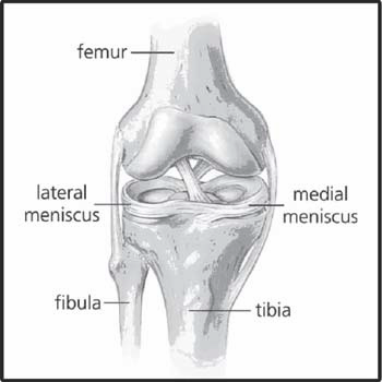 Most tears of the meniscus have limited healing ability, because the blood supply to the meniscus is limited to the peripheral 25-30 percent. Exceptions would include incomplete injuries to the outer border of the meniscus, especially in younger individuals. Although meniscus tears are not completely preventable, the best way to avoid meniscus tears is to have strong thighs and hips, along with flexible hamstrings.
Treatment for meniscal tears depends upon a person’s age, as well as the location, size, and chronicity of the tear. Incomplete tears in the peripheral border of the meniscus can often be managed with rehabilitation, short-term activity modification and anti-inflammatory medications. Nonoperative treatment includes standard R.I.C.E. therapy (Rest, Ice, Compression and Elevation). Occasionally, temporary crutch use is helpful to offload the stress on the knee. Anti-inflammatory medication use for a few days will decrease the pain that results from inflammation in the synovium or lining of the knee joint. Physical therapy can also be helpful to regain the strength and flexibility about the knee and hip. Steroid injections are helpful for nonoperative meniscus tears that are nonresponsive to a regimen of anti-inflammatories and therapy. Sometimes, getting over the acute flair of inflammation is all that is needed to get back to the pre-injury level of activity.
Meniscus injuries which block normal knee motion, causing persistent pain and functional limitations, are often best managed with knee arthroscopy – a minimally invasive technique for looking inside the knee joint. During arthroscopy, a physician places a small camera called anarthroscope into the knee to visualize any damage to the cartilage. Small instruments are then used to repair torn meniscus or shave down irreparable meniscus tears to a stable border so that the meniscus no longer catches in the knee. Following surgery, patients typically utilize crutches. Structured physical therapy,
together with a home exercise program, is often beneficial in facilitating your return to the court.
The following are common exercises that help with rehabilitation for both non-operative meniscus tears as well as following meniscal surgery. Start with the first 3 and move on to the second 3 when the pain has ceased.
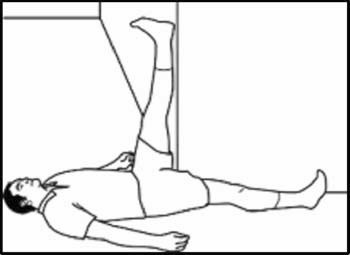
1) Hamstring Stretch: While lying on your back with your buttock close to a doorway, place your injured leg up on the wall. Slowly move your buttock closer to the wall. You will feel the stretch of the hamstring increase. Move to a comfortable stretch and hold for 30-60 seconds. Repeat 3 times.
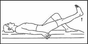 2) Straight Leg Raise: Sit on the floor with your uninjured leg bent and your injured leg straight. Press the back of your injured leg down, tightening the top part of your thigh (the quadriceps muscle). It is usually helpful to raise your toes toward your knee. Raise your leg about 6-8 inches from the ground and hold for 10-20 seconds. Repeat this 20 times. You can focus on different parts of your thigh by rotating your toes in or out.
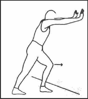 3. Calf Stretch: Face a wall and place your hands against the wall at shoulder level. Place your injured leg back and your uninjured leg forward. The uninjured leg should be slightly flexed and the injured leg should be straight with your foot flat on the ground. As you lean forward, you should feel the calf muscle stretch. Hold for 30 seconds. Repeat 3-5 times.
 4. Heel Slide: While sitting on the floor, slowly slide the heel of your injured knee toward your buttock, pulling your uninjured knee toward your chest. Return your leg to the extended position. Repeat 20 times.
5. Step-Ups: Stand with your injured leg on a block that is 3-5 inches high. Keep the foot of your uninjured leg flat on the floor. Shift your weight to the injured leg and straighten, lifting the uninjured leg off the floor. Return the uninjured leg to the floor. Repeat 10-20 times.
 6. Wall Squat: Stand with your back up against the wall. Keep your feet shoulder width apart and your feet 1-2 feet from the wall. Place a rolled-up towel or pillow between your legs. Squat down until your thighs are parallel to the floor. Hold for 20 seconds and slowly stand up. Keep the pillow or towel squeezed between your legs. Repeat 10-20 times.
Dr. Steve Hamilton, of Beacon Orthopaedics & Sports Medicine, is an orthopaedic surgeon who specializes in arthroscopic surgery. If your are suffering from pain or swelling of the knee, please schedule an appointment with Dr. Hamilton now or call (513) 354-3700 to schedule 24/7 via phone.
No Excuses – Ellen’s Story
September 27, 2017
Ellen came to us in June after an unfortunate slip on some steps. She had been training for a Triathlon in hopes to complete it and earn a medal that had her son in law and granddaughter featured on it. She wasn’t going to let a rotator cuff repair stand in her way! Read her words below. Great job Ellen!
“On June 29th Dr. McClung operated on my shoulder to repair a torn rotator cuff. At the time of the surgery I had already registered for a Triathlon in Muncie, Indiana. This Tri was very important to me as my son in law had taken first overall male winner in the past and as he crossed the finish line he held his daughter above his head as a celebration. Unbeknownst to him, a photographer captured that moment and the picture has appeared in magazines and on the web occasionally ever since. This year that picture was even the image used for the medal to be awarded for all distances at this event. Having participated in triathlons in the past, I was determined to earn that medal this year. Unfortunately, in the meantime I slipped on the stairs, injuring my shoulder and requiring the aforementioned surgery. Well, to make a long story short, just eight weeks after the surgery I did in fact get to participate. In case you don’t know. A triathlon in involves three stages – swim, bike, run. Per doctor’s orders I completed the 400 meter open water swim doing a one arm backstroke with my injured arm in a sling. To avoid stress on my arm, a fat tire bike with wide handle bars was used for the thirteen mile bike leg. A gentle 5K run wrapped it all up. Although it took some hard work and determination on my part, I couldn’t have done it without the care and encouragement I received at Beacon from Dr. McClung as well as my therapists.”
Is Ankle Replacement the Solution for You?
September 23, 2017
What is Total Ankle Arthroplasty (Ankle Replacement)?
Ankle replacement surgery serves to treat a patient with severe arthritis of the ankle by resurfacing the bone surfaces at the ankle joint. Metal components articulate with a plastic component to allow for pain relief and range of motion. Recent research has shown improved outcomes and survival rates for ankle replacements as a viable solution for severe ankle arthritis, and several patients at Beacon have taken advantage of this option with our team. Let’s go over some of the basic information you need to know about this exciting surgical treatment.
What is ankle arthritis?
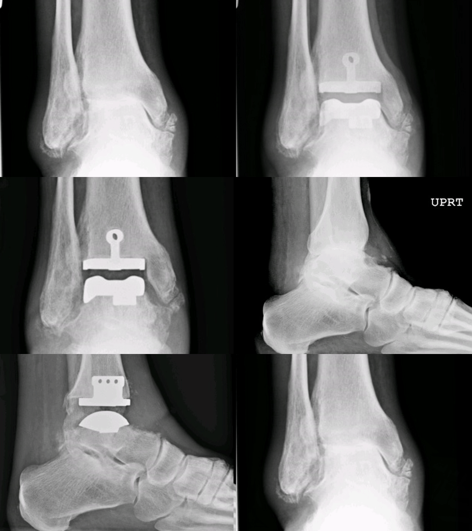 The ankle joint is a complex structure that includes three bones: the tibia (the major leg bone), the fibula (the smaller leg bone), and the talus. These bones interact to allow for motion at the ankle joint. Ankle arthritis is a condition in which the ankle joint cartilage wears out. Patients experience pain, loss of motion, swelling, and disability. There are several causes of ankle arthritis:
- Previous ankle trauma or fracture
- Ankle instability, severe ankle sprains over time
- Inflammatory arthritis (e.g. Rheumatoid)
- Osteoarthritis
- Other causes of arthritis such as gout or hemophilia.
What can be done to treat my ankle arthritis without surgery?
Nonsurgical treatment primarily consists of bracing and supporting the ankle to minimize painful motion. There are a variety of braces that can be used based upon the individual. Anti-inflammatory medicines are helpful for an acute exacerbation. Injection with corticosteroids on a limited basis may result in temporary relief of symptoms for pain. Although difficult, weight loss decreases the stress placed on the painful joint and typically relieves some pain.
If nonsurgical treatment fails, what can be expected with surgery?
Surgical options depend on the severity of the pain, amount of cartilage damage, and patient’s occupation, age, weight, and activity level. Early arthritis may benefit from an isolated removal of large bone spurs if present, with or without arthroscopy. The majority of ankle arthritis is treated by one of two methods: ankle fusion or ankle replacement. This is a complicated surgery that requires an orthopaedic surgeon’s background and experience in ankle replacement. The risks and benefits of each treatment can be complicated and are better suited for a full discussion with our team after a full clinical and radiographic evaluation.
What happens after surgery and how long will this affect my walking?
Ankle replacement surgery can be performed as an outpatient or an overnight stay at a hospital. A nerve block will help control pain during and after surgery. The leg would be protected in a splint until seen in the office 8-10 days later. A boot is used as a cast until seen 6 weeks after surgery. There is a period of no weight bearing initially after surgery to allow the bones to heal to the implants. Recovery usually occurs progressively over a six-month period.
Appointments can be made with Beacon’s foot & ankle specialist Dr. Adam G. Miller by calling (513)-354-3700 or booking online here.
Recovery Guide to Foot & Ankle Surgery
September 12, 2017
We are glad that you have chosen Dr. Miller for your foot and ankle surgery. Our goal is to make this experience as pleasant as possible for you. If you have any questions about your hospital stay, rehabilitation or recovery period, please contact Dr. Miller’s office for more information specific to your surgery.
Please click here to download the full informational PDF guide.
The Difference Between Herniated and Bulging Discs, and What That Means for You
September 6, 2017
Back pain is the second most common reason people take sick days—second only to the common cold. That statistic accounts for workers from every industry, indicating that back pain is not limited to any single job type. 70-85% of all people experience back pain severe enough that it requires treatment at least once in their lives, and back pain is the leading cause of disability worldwide.
Two of the most common causes of back pain are obesity, and improper technique when lifting heavy objects. Worldwide obesity rates have more than doubled since 1980. The spine and surrounding muscles simply cannot support the extra weight an obese person carries, and can result in a variety of spine injuries. Using your back muscles to lift a heavy object instead of your legs is improper lifting technique, and often results in a back injury. Compared to your leg muscles group (gluteus maximus, quadriceps, hamstrings, etc.) your lower back specifically, is much weaker and likely to become injured.
Some of the most common types of back injuries besides strained and pulled muscles, are herniated discs, also called slipped discs or ruptured discs. They are often confused with bulging discs, which are also very common and similar. This article will help you understand the difference between a herniated disc and a slipped disc, risk factors and treatment options for these types of injuries.
The Anatomy of Your Spine
Without a healthy spine, you wouldn’t be able to move. While the brain is in charge of signaling your body what to do, the spine is in charge of relaying those signals to the right places. Without the spine, the brain would be like an engine without a car to power.

The spine is made up of bones called vertebrae that protect the delicate spinal cord. Between those vertebrae are small, round cushions made of fibrocartilage and collagen, called vertebral discs. Each vertebral disc has a soft inner nucleus, consisting of a gel-like substance called the nucleus pulpous, and is surrounded by a tough, fibrous wall. The vertebral discs allow the spine to bend and twist, and act as a shock absorber, preventing vertebrae from compacting on one another.
What is a Herniated Disc?
Herniated discs get their name from hernias, which occur when an organ pushes through the muscle or tissue that holds it in place. These most commonly occur with the intestines and the abdominal wall. The same concept applies to a herniated disc, the gel-like fluid in the center of the disc pushes through the fibrous outer wall of the disc. This herniation of the disc can result in a large bulge that can press on nearby nerve roots, causing pain.

However, herniated discs don’t always hurt. In fact, it’s entirely possible to have a herniated disc and not know it. Herniated discs only cause pain when the herniated area pushes on a nerve root in the spine. Pain from a herniated disc can occur at the vertebral disc where the herniation has occurred and/or in other places, like the legs or arms. The location of pain depends on what the affected nerve root is connected to. In extreme cases, a herniated disc will cause your arms or legs to tingle, or even go numb due to the pressure on the affected nerve. In these cases, you should call 911 immediately.
What is a Bulging Disc?
A bulging disc occurs when the fibrous outer wall of the disc weakens and the nucleus pulpous is pushing to get out, but has not yet ruptured through. This causes the disc to bulge outward, which is where the condition gets its name. Bulging discs are less likely to cause pain than herniated discs because they generally do not protrude far enough to press on a nerve. However, a bulging disc is often progresses into full-blown herniated disc over time.

Clarifying the Difference
Talking about herniated and bulging discs often gets confusing because of how similar they are. That’s because the term “herniated disc” actually refers to a disc in one of four stages of herniation:
- Degeneration (this is when it bulges initially)
- Prolapse (this is when it bulges more)
- Extrusion (this is when the gel actually slips out of the disc)
- Sequestration (this is where the gel is no longer attached to the disc)
A disc in the degeneration or prolapse stage (what we call a bulging disc) is technically still a herniated disc, even though the herniation has not yet occurred. This accounts for basically all of the confusion surrounding herniated and bulging discs. The term “slipped disc” is used to refer to a herniated disc as well, because the nucleous pulposus has “slipped” out of place.
So, when someone talks about a bulging disc, they are referring to a disc in either the degeneration or prolapse stage. When someone talks about a herniated disc, they could be referring to a disc in any of the four stages. It’s impossible to know which exactly they mean unless you know for certain that they understand the difference between a herniated and a bulging disc.
What are the Risk Factors for a Herniated Disc?
- Aging: Past age 30, spinal discs gradually lose fluid volume. This causes them to dry out and become more brittle. This process cannot be reverted.
- Improper lifting mechanics: Whether you’re a weight lifter or just helping a friend move, lifting objects with a curved back puts extreme pressure on your spine. When you lift, do so from a squatted position, with your knees bent and your back straight. For more information on proper lifting form, check out our blog post on the subject.
- Repetitive stress: If you do something that stresses your lower back frequently, like swinging a golf club or a baseball bat for example, your risk of herniating a disc is increased.
- Obesity: Obesity puts you at high risk for many types of health problems, and vertebral disc issues are no exception. As you gain weight, you put more and more pressure on your discs, increasing the risk of rupture.
- Genetics: Disc health has a genetic component to it, and if your family has a history of herniated discs, it’s more likely that you will incur them, too.
- Traumatic Injuries: It’s rare, but occasionally an injury, such as a nasty fall, car accident, etc., can put so much stress on a disc at once that it herniates.
How are Herniated Discs Diagnosed?
When a patient comes in with back pain, a physician will usually take three steps to determine whether or not the patient has a herniated disc:
- The physician will consider the patient’s medical history, since poor vertebral disc health can be inherited. They will also have the patient describe when the pain started and where the pain occurs.
- Next, the physician will do a physical exam. They will test nerve function and muscle strength in the arms and legs. Most of the time, a physical examination tells a doctor everything he needs to know about the nature of an injury.
- If necessary, the physician will order a CT or MRI scan on the patient. Bulging or herniated discs are easy to spot with such scans.
How are Herniated Discs Treated?
Depending on the severity of the herniated disc, there are three possible treatment options available:
-
Pain management
Pain management seeks to minimize the discomfort herniated discs cause. This is done either through medicine or injections. While these treatments are effective at reducing pain, they do nothing to stop the herniation from pressing on the spinal nerve.
-
Physical therapy
Physical therapy is a great treatment option for herniated discs because it can both relieve pain and reduce the likelihood of incurring future disc-related injuries. Poor posture, weak muscles, and obesity all contribute to a disc herniating, and physical therapy helps improve all of the above.
-
Surgery
Herniated discs can almost always be treated without surgery, but surgical treatments are also an option, if necessary. The orthopedic spine surgeon removes the herniated section of the disc, and closes the disc back up, effectively treating both the herniated disc and the pain associated with it.
Beacon Can Get You Back on Track
Whether you’re currently experiencing back pain or are just worried that your posture will give you trouble in the future, Beacon Orthopaedics and Sports Medicine offers a wide variety of treatment options that can help you. Our resident spine expert, Dr. Michael Rohmiller, is a master of minimally invasive spine surgery and can work with you to find the best treatment option. We also have an excellent team of physical therapists who can tailor a plan to address your disc problems pre or post herniated disc. Schedule a meeting with Dr. Rohmiller today to discuss your injury and treatment options.
Know When You Need Surgery: Conditions Treated by Knee Replacements
August 28, 2017
Most people will experience some degree of knee pain or a knee injury in their lifetime, and often those injuries are minor and may heal on their own. However, many people experience consistent knee pain for years, often because of an untreated injury, arthritis or other degenerative diseases. For many older patients experiencing intense knee pain, a partial or total knee replacement can be an excellent treatment option.
Beacon Orthopaedics has three steps to help you decide when it’s time to consider a partial or total knee replacement, and the specialists who can help you get the treatment you need.
1. Determine the Severity of Your Condition/Knee Pain
As you weigh your options for pain management and treatment, ask yourself the following questions:
- How much does my knee hurt in motion?
- How much does my knee hurt at rest?
- What activities make my pain worse?
- Do I experience dull, mild, or severe pain?
- Do I experience consistent pain?
- How much can I move my knee before I experience pain?
- How often does my knee hurt?
- Is there swelling? How much?
2. Identify the Cause of Your Knee Pain
The best way to identify the cause of your knee pain is to see an orthopaedic specialist. Below are some of the most common causes of knee pain in order of their severity:
- Excessive pressure on the joints
- Ligament strains, sprains and tears
- Tendonitis and bursitis (inflammation due to overuse)
- Fractures and breaks
- Dislocation of the joint
- Gout (crystalized uric acid in a joint)
- Post-traumatic arthritis (cartilage damage due to injury)
- Rheumatoid arthritis (excessive synovial fluid in the joint)
- Osteoarthritis (wearing down of cartilage in the joint)
3. Consider How Your Symptoms Affect Your Everyday Activities
The majority of people will experience knee pain at some point during their life. Even the smallest fall or slightest twist can cause lasting damage to the knee. When your symptoms interfere with the quality of your everyday life, it’s imperative that you consult a knee specialist.
When Should I See a Specialist for a Knee Replacement?
Consider talking to a knee specialist if it is difficult to perform any of the following activities without pain:
- Walking
- Climbing stairs
- Getting in/out of your car
- Getting in/out of bed
- Getting in/out of the shower
- Getting on/off the toilet
Also consider talking to a knee specialist if you consistently experience:
- An inability to bear weight on your knee without pain
- Severe pain, swelling, instability, or grinding in your knee
- Persistent pain or swelling that does not resolve in a few weeks
- Pain or stiffness that limits your everyday activities, including walking, climbing stairs, and getting in and out of chairs
- Moderate or severe knee pain while resting or sleeping
- A knee deformity – such as the bowing in or out of your knee
Common Conditions Treated by Knee Replacement
The most common cause of chronic knee pain, and a condition that will often result in the need for knee replacement surgery, is arthritis. The three forms or arthritis that primarily affect the knee are:
Post-traumatic Arthritis – Usually following a serious knee injury, this type of arthritis is caused by the gradual damage to articular cartilage from fractures in the bones surrounding the knee, or tears of the knee ligaments.
Rheumatoid Arthritis – This type of arthritis is caused by the inflammation of the synovial membrane that surrounds the knee joint, the resulting chronic inflammation can damage the cartilage and eventually cause cartilage loss, pain, and stiffness in the knee.
Osteoarthritis – This is an age-related type of arthritis that is directly caused by the wear and tear of the knee joint over time. It usually occurs in people 50 years of age and older, but may occur in younger people, too. As the cartilage that cushions the knee softens and wears away, the bones then rub against one another, causing knee pain and stiffness.
While post-traumatic arthritis, rheumatoid arthritis, and osteoarthritis have different causes, they all cause loss of cartilage in the knee. When the knee no longer has enough cartilage to cushion the movement of the leg, walking becomes painful. It is imperative to speak to a specialist about a knee replacement in order to avoid further damage to the bones in the knee.
Here is additional information about arthritis and knee replacement that will help guide your discussion with your physician.
Benefits to Knee Replacement Surgery
- Ease Pain in the Knee
- Regain and Preserve Overall Mobility
- Arthritic Bone is Completely Removed
- Correct Knee Deformities
Beacon is a Leader in Knee Replacements
The goal of total knee replacements is to restore function, stability, and alignment in the knee while relieving pain in the process. In order to fully achieve this, specialized experience and support is required.
If you experience knee pain that interferes with daily living, schedule an appointment with Dr. Haleem Chaudhary at Beacon Orthopaedics and Sports Medicine to talk about possible causes and treatments. Dr. Chaudhary is board certified and fellowship trained in adult joint reconstruction. He will guide you from your pre-operative consultation all the way through recovery, ensuring that your new knee will fit comfortably and provide you with the support you need to live your life to the fullest.
Learn more about Dr. Chaudhary or schedule an appointment to meet with him at Beacon East, Summit Woods, and Beacon West in Ohio or at Beacon’s Northern Kentucky location.
The Benefits and Risks of Tommy John Surgery
August 15, 2017
A professional baseball pitcher throws tens of thousands of full-force pitches over the course of their career. These athletes depend on their ulnar collateral ligament (UCL), a tiny band of tissue located in the elbow, in order to repeatedly and consistently pitch at speeds of 90 mph or more. Over time however, the force generated by their throw and the repetitive stress of pitching can cause a UCL to tear. A UCL tear inhibits a pitcher’s ability to grip a ball, perform overhead motions, and throw with the speed necessary to compete.
Fortunately, UCL tears are no longer the career ending injuries they once were. Ulnar collateral ligament reconstruction—better known as Tommy John surgery after the Dodgers pitcher who first received the surgery in 1974—can effectively treat a tear. During the surgery, the damaged UCL is completely replaced with a healthy tendon, providing effective relief of the patient’s symptoms. However, Tommy John surgery is not always necessary for UCL injuries, and like all surgical procedures, there are risks of complications. It’s important to know the benefits and risks of Tommy John surgery in order to guide your discussion with an orthopaedic specialist.
What Leads to Tommy John surgery?
In order to understand the benefits of Tommy John surgery, it’s important to first understand the symptoms that the surgery is intended to treat.
Anatomy of the Elbow
The ulnar collateral ligament connects the humerus (upper arm bone) to the ulna (forearm bone opposite of the thumb). The UCL stabilizes the inside of the elbow against valgus stress, or stress that results from the hand moving away from the body. While the UCL can handle a reasonable amount of stress, force that exceeds its’ tensile strength can stretch or tear the ligament. Moreover, repetitive use of the elbow can cause microscopic, undetected tears to develop. These tears gradually increase in size, weakening the ligament and consequently making it more susceptible to further injury.
UCL tears are common among athletes who make frequent overhand motions, including javelin throwers as well as football, tennis, and hockey players. Professional baseball pitchers – by far – face the highest risk of a UCL tear. In fact, approximately 1 in 4 injuries sustained by professional pitchers is related to the elbow. This is due in part to the amount of valgus stress generated by the average pitch. During the acceleration phase of an overhead throw, the forearm lags behind the upper arm. This puts stress on the elbow that can exceed 60 newton meters (N m), which far exceeds the 34 N m the ligament can handle. In short, every full-force pitch that a pitcher throws poses a serious risk of a tear.
When a tear develops, an individual may experience:
- Pain on the inside of the elbow
- Pain when using the arm in an overhead position
- Swelling along the inside of the elbow
- Decreased hand grip and forearm strength
- Instability in the elbow
While muscles around the elbow can be strengthened in order to compensate for this instability, athletes often need to do more than just manage the symptoms of a tear—they need to treat the condition at its source. An orthopaedic specialist may recommend Tommy John surgery in order to restore normal elbow function and help them return to their sport.
How the Surgery is Performed
Tommy John surgery is performed by using a grafted tendon to replace the damaged ligament. The surgery begins with an incision on the inside of the elbow joint. Then a healthy tendon is harvested from another area of the patient’s body or a donated tendon is prepared. Next, the damaged ligament is removed and replaced. Finally, the incision is stitched up and the elbow is placed in a large bandage. A typical Tommy John procedure takes 90 minutes. Because the surgery is an outpatient procedure, patients can also return home the same day.
The Benefits & Risks of Tommy John
Tommy John surgery relieves pain and restores proper elbow function. The new UCL ligament is healthy, strong, and allows the patient proper use of the elbow. Post-surgery, the patient must re-build their strength via months of physical therapy and rehabilitation, which could last anywhere from 6 months to a year.
While Tommy John surgery has a high rate of success in allowing pitchers return to the mound, not every athlete returns to his or her pre-injury level of performance. There is also always the risk of re-injury post-surgery if the proper rest and recovery programs are not followed.
Prevention
While Tommy John surgery has never been more widely performed than it is today, it is a difficult and highly technical operation. UCL injuries are extremely preventable for baseball players by properly warming up, maintaining proper form, and following proper pitch counts and rest day recommendations based on age. Tommy John Surgery is always the last resort and will be considered only after all other conservative treatment options have been exhausted. It’s imperative that pitchers focus on proper body mechanics at all times in order to both preserve the health of their elbow and avoid additional surgeries.
Talk to an Orthopaedic Specialist
When Tommy John underwent the first ulnar collateral ligament reconstruction in 1974, it was estimated that he had a “one in 100” chance of returning to baseball. Today, approximately 85 percent of pitchers who receive Tommy John surgery return to the mound after a year of recovery.
Dr. Timothy Kremchek at Beacon Orthopaedics and Sports Medicine is a board certified orthopaedic surgeon who is fellowship trained in sports medicine and one of the premiere Tommy John surgeons in the nation. Dr. Kremchek is the Team Physician for the Cincinnati Reds and has worked with hundreds of professional, collegiate and high school athletes. With over 1,500 Tommy John surgeries performed, he is uniquely qualified to talk to you about your condition and answer any questions you may have regarding the procedure. You can schedule an appointment online to meet with Dr. Kremchek at Beacon’s Summit Woods or Wilmington locations.
Low Vitamin D: Are Your Bones At Risk?
August 3, 2017
The Need for Vitamin “D”:
Vitamin D has been a hot button topic for years now. Why is it so important? Why do so many people have low levels and what are the implications of that?

The more we learn, it has become apparent the importance of this metabolite. Low “D” has been implicated in conditions such as rickets and osteoporosis. In fact, milk and other products were mandated fortification with vitamin D in the 1930’s due to rickets and the associated bone malformations observed in children. Besides consumption of dairy and now some orange juice, some fish obtain high levels of the vitamin.
The requirement of vitamin D in the diet comes from the decreased sun exposure that nearly all of us see. Whether you blame increased work of adults or video games in children, less outside activity is prevalent. In addition, dermatology places strong emphasis now on protection from skin cancers and melanoma through the use of sun block. The prevention of ultraviolet sun exposure to skin also prevents the activation of the metabolite.

Can Low “D” Cause Injury?
Beyond rickets (which is rare today with fortified products) and osteoporosis, vitamin D is associated with increased chance of injury. Recently a common fracture to the 5th metatarsal called a Jones fracture was recently found to be five times more likely in patients with low levels of “D”. There is also a suggestion that “bone edema” or bruising of bones is associated with low vitamin D levels. This suggests that elevating a low level of the metabolite may prevent some of these fractures. Therefore replenishment has been suggested to be appropriate is most cases if detected.
A normal vitamin D has been determined to be above 30ng/mL. Low vitamin D is highly prevalent in latitudes above 30 degrees (North of Jacksonville, Florida). So, chances are you have low levels! Before every surgery involving bone, I check a patient’s vitamin D level to ensure there is a minimal chance this will effect outcomes. Next time your doctor requests blood work, it may be worth checking…
For more information or a personal evaluation, please visit our website. Appointments can be made with Dr. Adam G. Miller by calling (513)-354-3700 or booking online here. If you are not following Dr. Adam G. Miller on social media, you can do so on Facebook or Twitter for: updates and comments on cutting edge treatments, discussion of various injuries, and sport/athlete issues.
Frequently Asked Questions about Rotator Cuff Injuries
July 17, 2017
What is a rotator cuff injury?
The shoulder consists of the humerus (upper arm bone), scapula (shoulder blade), and clavicle (collarbone). The rotator cuff is a group of four muscles that hold the head of the humerus and scapula together. While its primary function is to help stabilize the shoulder, it also aids in rotational movement. Injuries occur when any of these structures have been damaged.
What are the most common types of rotator cuff injuries?
Tendinitis, bursitis, and tears are the three most common types of rotator cuff injuries.
Rotator cuff tendinitis, also called shoulder impingement syndrome, occurs when the tendons that connect the rotator cuff muscles to the humerus become irritated or inflamed. Similarly, bursitis occurs when bursae, or the fluid-filled pads that serve as cushions in joints, become painful and swollen. These conditions share a number of causes, including:
- Repetitive overhead movements
- Holding the arm in the same position for long periods of time
- Compressing the arm for long periods of time
- Sudden injury
- Osteoarthritis
Rotator cuff muscles and tendons can also be partially or completely torn. A tendon that has been partially frayed, or worn, is classified as a partial-thickness tear. By contrast, full-thickness tears range from pinpoint holes in the muscles and tendons of the rotator cuff to substantial tissue damage. This type of tear can affect one or more tendon and, in the most severe instances, completely detach rotator cuff tendons from the humeral head.
Moreover, tears are categorized as either acute tears or chronic tears depending on their cause. Acute tears result from sudden injuries such as falling on an outstretched arm or lifting a load that is too heavy. Chronic tears, also known as degenerative tears, often begin as microscopic injuries and gradually worsen over time.
What are the signs and symptoms of a rotator cuff tear?
Individuals may experience a sudden “tearing” sound and/or sensation at the time of their injury, followed by weakness in the affected arm. Other patient have a gradual decrease in shoulder motion and strength from a chronic degenerative tear.
Symptoms of a rotator cuff tear include:
- Shoulder pain
- Loss of shoulder mobility
- Weakness in the shoulder joint
- Swelling around the shoulder
- Pain with overhead activity
While symptoms can vary from mild to severe, it’s important to understand that the amount of pain associated with a tear does not necessarily correlate with its size. In fact, patients with a small, partial tear—or even no tears at all in the case of tendinitis and bursitis—can easily mistake their pain as a sign of a worse condition. Conversely, major tears may only be mildly painful and even go unnoticed. The same principle also applies to shoulder mobility.
Only an orthopaedic specialist can properly diagnose a rotator cuff tear. If you suspect an injury, it’s important to see an orthopaedic specialist as soon as possible.
What are the risk factors for a rotator cuff tear?
Overuse: Repetitive, overhead motions associated with certain sports and occupations put individuals at greater risk of chronic tears. This type of injury is particularly common in overhead sports such as: baseball, tennis, golf, volleyball, and swimming. Moreover, construction workers and those who work in similar, manual labor type fields are also at high risk of developing a chronic tear.
Arthritis: Degenerative forms of arthritis—especially osteoarthritis—will weaken rotator cuff muscles and tendons, consequently making them more susceptible to tears.
Age: Individuals over the age of 40 are more likely to develop rotator cuff tear. This is primarily due to two natural changes the body undergoes as it ages. First, less blood is supplied to the rotator cuff tendons. This diminishes the body’s ability to heal these tendons and consequently makes them more susceptible to injury. Second, bone spurs often develop on the underside of the acromion which as located at the tip of the shoulder blade. These spurs, or overgrowths, rub on rotator cuff tendons every time the arm is lifted. Over time, this can not only lead to shoulder tendinitis, but also weaken the tendon and lead to a potential tear.
How is a rotator cuff tears diagnosed?
The diagnostic process begins with a patient interview and is followed a physical examination. During the examination, the orthopaedic specialist will:
- Press on different parts of the shoulder
- Maneuver the shoulder through various positions
- Test the strength of shoulder and arm muscles
Often, a physical examination is all that is needed to diagnose a rotator cuff tear. However, a specialist may recommend imaging tests such as x-rays, ultrasound, or an MRI to help guide treatment options and confirm the size or location of the tear.
What treatments are available for rotator cuff tears?
Most rotator cuff tears can be treated with a combination of rest and physical therapy. And while conservative, non-surgical treatments can be highly effective, patients will need to demonstrate patience. Moreover, patients will need to protect their shoulder against further wear and tear, as well as additional complications, while their body heals.
Patient who do not respond to the initial nonoperative program may have their symptoms improve with orthobiologics or surgical intervention. orthobiologics treatment involves injections to stimulate the body’s ability to heal itself. Surgery involves arthroscopic techniques that allow the surgeon to remove bone spurs, bursitis and inflammation. This minimally invasive technique also allows the surgeon to repair the torn tissue if needed.
The most severe rotator cuff injury, a large full-thickness rotator cuff tear, typically requires surgery. Arthroscopic rotator cuff repair, in particular, is a minimally invasive surgery that an orthopaedist may recommend to younger patients who are in otherwise good health. This form of surgery allows a surgeon to repair the patient’s damaged muscle and tendons while also minimizing overall tissue trauma.
What is the outlook on recovery?
Minor to moderate rotator cuff tears require approximately 6 to 12 weeks of rehabilitation. Physical therapy not only helps restore shoulder strength and mobility but it also prevents the build-up of scar tissue around the shoulder joint. While many patients will recover most of their shoulder strength and mobility, some patients will never fully recover.
While large rotator cuff tears are uncommon, a combination of conservative treatments and surgery has been proven to be highly effective. If left untreated, a large tear can lead to arthritis. Specifically, the combination of a rotator cuff tear and arthritis is referred to as rotator cuff arthropathy. Unfortunately, there are limited options for this condition. In the majority of cases, a shoulder replacement is necessary to restore shoulder strength, mobility, and function. It’s imperative that patients see an orthopaedic specialist immediately following an injury in order to avoid arthropathy and similar complications.
Additional Resources
Beacon Orthopaedics and Sports Medicine offers a variety of educational material related to rotator cuff tears. The following are related articles and videos from our blog:
If you are experiencing shoulder pain, and conservative treatment options haven’t helped, schedule an appointment with Dr. Steve Hamilton for an evaluation and treatment plan personalized to you.
Preventing Shoulder Cuff Injuries in Athletes
July 7, 2017
The shoulder has the greatest range of motion of any joint in the body. It allows the arm to move away from the body’s midline, towards the body’s midline, forward, and backwards. It can also move the arm in a full circle, as well as rotate it towards or away from the midline. The articulation of the shoulder is so unique, in fact, that the hip is the only other joint in the human body that is also classified as a spheroidal joint, which is commonly known as a “ball and socket” joint.
While the rotator cuff is highly complex, it is also relatively weak. Similar to other structures of the body, it can be damaged by traumatic injuries, and is also extremely susceptible to wear and tear. Athletes who excessively use their shoulder—such as baseball and tennis players—are at the greatest risk of a rotator cuff injury. But, as any of these athletes would tell you, repetitive use of the shoulder is an unavoidable part of the game.
So what can athletes do to prevent rotator cuff injuries? Like all other sports-related injuries, prevention entails education, physical conditioning, and practice.
The Anatomy of the Rotator Cuff
The shoulder consists of the humerus (upper arm bone), the scapula (the shoulder blade), and the clavicle (the collar bone). The joint is formed from the spherical head of the humerus, which acts as a “ball”, and the glenoid cavity, which forms the “socket”.
The rotator cuff is a group of four muscles that come together at the head of the humerus. These muscles not only help stabilize the shoulder but they also keep the arm in its socket. In addition, each muscle also serves a specific, unique function related to movement.
- Infraspinatus: Rotates the arm laterally (away from the midline) at the shoulder joint.
- Supraspinatus: Helps the deltoid muscle abduct (raise) the arm at the shoulder joint.
- Subscapularis: Rotates the arm medially (towards the midline) at the shoulder joint.
- Teres minor: Rotates the arm laterally and weakly adducts (lowers) the arm at the shoulder joint.
Common Rotator Cuff Injuries
Rotator Cuff Tears
Rotator cuff tears are a common orthopaedic condition. When a rotator cuff muscle tears, it partially or completely detaches from the head of the humerus, causing shoulder pain and instability. Tears are most likely to occur in the supraspinatus muscle and tendon. Because these injuries result from a variety of causes and range in severity, they can be categorized in a number of ways.
Acute Tears
An acute tear results from a sudden injury, such as blunt force to the shoulder or a fall on an outstretched arm; however, a rotator cuff with pre-existing degeneration can also be torn by simple, everyday activities such as gardening or putting away the dishes.
Chronic Tears
A chronic tear, also called a degenerative tear, results from repetitive use of the shoulder cuff muscles over time. Athletes are at especially high risk for this type of overuse injury. Degradation, like the kind that is associated with chronic tears, occurs naturally with age as well. As such, people over 40 are at increased risk for this type of injury. Some common causes include:
- Reduced blood flow – Blood oxygen levels naturally decrease as people become older. Since oxygen is essential to all parts of the body, reduced oxygen both increases the risk of a rotator cuff tear and makes healing more difficult.
- Bone spurs – Bone spurs are little growths that occur on the bone as a result of too much calcium. These spurs rub on the rotator cuff, causing pain and degradation. Bone spurs can also lead to shoulder impingement, a condition where the arms cannot go above shoulder level without severe pain.
Signs of a rotator cuff tear include:
- A sudden tearing sensation
- Immediate weakness in your arm
- A cracking sensation when moving your arm
- Pain during certain movements
Symptoms of a rotator cuff tear include:
- Shoulder pain that gradually worsens over time and persists throughout the night
- Shoulder weakness that gradually worsens over time
- Difficulty performing overhead movements
A rotator cuff tear is described as either a partial or full thickness tear based on its severity.
Partial Rotator Cuff Tears
This form of injury is also referred to as partial thickness rotator cuff tear because the tear only extends part way through the tendon and often only involves one of the four rotator cuff muscles. It is more common than a complete tear and more common in younger individuals. And while seemingly less severe than a complete tear, there is no correlation between the size of a tear and the amount of pain it causes. In fact, in some instances, a partial tear can be more painful than a full-thickness tear.
Full-Thickness Rotator Cuff Tears
A full-thickness rotator cuff tear occurs when one or more muscles and tendons completely separate from the humerus. Large tears can cause significant shoulder weakness. For example, an individual may have to support their injured arm with their other arm when lifting or moving an object. Large tears can also cause a loss of shoulder mobility; however, this is not always the case.
Tendinitis and Bursitis
Tendinitis causes tendons to become swollen and painful, often due to over use and inflammation. Similarly, bursitis occurs when bursae—small fluid-filled sacs between bones, tendons, and muscles—become inflamed. These conditions can occur alone or simultaneously and are more likely to occur in people age 40 or older.
Tendonitis, in particular, is often a precursor to a tear. When tendons become inflamed, they scrape against the bones in the shoulder joint. This frays the tendons and consequently makes them more susceptible to tears.
Treatments for Rotator Cuff Injuries
First Aid
RICE (Rest, Ice, Compression, and Elevation) should be applied as soon as possible after an injury occurs. Rest provides time for the injury to heal while ice and compression reduce the amount the amount of swelling. In the case of rotator cuffs, however, individuals may forgo elevating the arm if it is difficult or uncomfortable.
Professional Treatment
The majority of rotator cuff injuries can be treated with a combination of rest and physical therapy. Physical therapy helps restore shoulder flexibility and strength as well as reduce pain.
If a patient does not achieve relief with conservative treatments, a physician may recommend orthobiologics, which is centered on the body’s own ability to itself. orthobiologics aids the body in creating new, functional tissue to replace old and damaged tissue.
Surgery is also an effective option; however, it is often a method of last resort. Candidates for surgery have symptoms that limit normal, everyday function and do not improve with conservative treatments. A talk with an orthopaedic specialist can determine if surgery is the right option for you.
How to Prevent Rotator Cuff Injuries
In general, the best way to prevent rotator cuff injuries is to strengthen the rotator cuff. Keep in mind that even simple, daily exercise can go far in strengthening muscles and tendons. The “doorway stretch” is one such exercise that can be performed at home. To perform the exercise:
- Stand in an open doorway and spread your arms out to your side.
- Grip the sides of the doorway at shoulder height.
- While maintaining your grip and keeping a straight back, lean forward until you feel a light stretch in the front of your shoulder.
- Slowly return to the original position. Repeat 10 times.
As always, speak with a physical therapist before starting an exercise program.
In addition to exercise, athletes should focus on proper form. Improper form puts increased stress on the rotator cuff and increases an athlete’s risk of an injury. Frequent breaks can help an athlete avoid fatigue that consequently leads to improper form.
When to See an Orthopaedic Specialist
While the majority of rotator cuff injuries heal with rest, it’s a slow process. Moreover, because many everyday activities require the use of the shoulder, patients can potentially worsen their condition by not seeking early treatment. For all of these reasons, it’s important to talk with an orthopaedic specialist at the first sign of an injury. An orthopaedist will ensure that you’re on the best path to recovery.
Dr. Robert Rolf is a board certified orthopaedic surgeon at Beacon Orthopaedics and Sports Medicine who provides extensive expertise in rotator cuff tears as well as other conditions related to the shoulder or elbow. Patients can meet with Dr. Rolf at Beacon’s Batesville, Lawrenceburg, or Northern Kentucky location as well as Beacon West in Harrison, Ohio. Dr. Rolf also offers informational Shoulder Talks at Beacon West. For a list of upcoming talks, visit Dr. Rolf’s page.
Proper Lifting Technique to Avoid Back Injuries
July 5, 2017
More than one million injuries are sustained in the workplace each year. Back injuries, in particular, account for 1 in 5 of these injuries and is the single leading cause of disability worldwide.
At Beacon Orthopaedics and Sports Medicine, we frequently treat patients suffering from occupational back pain. While some work-related back pain can be attributed to poor posture, the majority of injuries are caused by improper lifting technique. The truth of the matter is that lifting even a light load off the ground puts more strain on the body than many people realize. Simply put, there is no such thing as a “simple” lift. Any time an object needs to be moved, it requires proper technique.
Here are the steps to follow in order to avoid back injuries while lifting a heavy load.
Proper Lifting Technique
1. Evaluate the Load and the Surrounding Environment
First and foremost, determine if the load can be safely handled. Do not attempt to lift a load if it appears too heavy or difficult to handle. The surrounding environment should allow for proper footing and be free of potential tripping hazards.
2. Position Your Body Close to the Load
Position your body close to the load with your feet shoulder width apart and one foot slightly ahead of you.
3. Squat Down with Your Back Fully Straight
Squat down close to the load. If possible, squat until you’re “below parallel”, meaning your hip joint is at or below the level of the knee joint. Many individuals only squat until their hip is parallel with their knees, creating a 90 degree angle or an L-shape; however, research suggests that dropping below parallel activates the larger muscles in the lower body. When the body is able to rely on these larger muscles, less stress is placed on the other areas of the body.
Also ensure that your back is fully straight and not just vertical. To keep your back straight, move your shoulders back and push your chest out more. You can also tuck in your chin to help straighten your back. When lifting a large load, you may bend at the waist in order to keep the load close to your body.
4. Securely Grip the Load
Grip the load, keeping it close to your body. At this point, if you are still certain that you can safely handle the load and maintain a straight back, you can proceed.
5. Slowly Lift the Load with Your Legs
Use your body weight to initially lift the load from the ground and then continue to lift by straightening your hip and pushing with your legs. As you lift, avoid twisting your torso. If you must change direction, turn your body by taking small steps and leading with your hips. Moreover, it’s important to maintain a straight back the entire time. Bending the back moves the load away from the body. The resulting leverage increases the stress on the lower spine and nearby muscles.
A lift should end with your legs fully straightened and the object resting between mid-thigh and shoulder height. Holding an object above shoulder height puts stress on the upper back, shoulders, and arms.
6. Set Down the Load
Slowly squat in order to lower yourself and the load to the ground.
Dangers of Poor Lifting Technique
The back is one of the most complicated regions of the body. Broadly speaking, the back consists of two major muscles groups; an interlocking series of bones referred to as vertebrae; a series of intervertebral discs that separate and cushion these bones; multiple facet joints that provide the back with movement; numerous ligaments that connect and stabilize the spine; and, most importantly, the spinal cord. Poor technique can harm any and all of these structures.
Muscle Injury
The majority of injuries caused by improper lifting techniques are either muscle strains or ligament sprains in the lower back. A muscle strain occurs when weak muscles have been overstretched or torn. A ligament sprain, on the other hand, occurs when the fibrous tissue between bones has been stretched or torn. While seemingly simple injuries, both strains and sprains can cause severe lower back pain.
Disc Injury
Discs are located between the individual vertebrae of the spine and function as shock absorbers. Poor technique can cause vertebral discs to shift out of alignment. When this happens, the discs can come in contact with root nerves or the spinal cord, causing tremendous pain. Discs are also at risk of rupturing or, in other words, breaking open. A ruptured disc, otherwise known as a herniated disc, occurs when a crack develops in the wall of a disc and its inner contents are pushed out into the spinal canal. Similar to a bulging disc, a ruptured disc is painful and requires treatment from an orthopaedic specialist.
Joint Injury
Each section of the joint contains facet joints which are what provide the spine with its flexibility. When these joints have been injured, they stiffen. Many patients describe this stiffness as “buckling” or “locking up.” An injured back joint will also cause lower back pain and potentially refer pain to the buttock or thigh.
The Role of Physical Conditioning
Strengthening
While the majority of lifting injuries result from improper technique, some are simply the result of trying to lift a load that is too heavy. In order to avoid these injuries, individuals should know how much they can safely lift. If an object seems too heavy, ask for help. If lifting heavy loads is a necessary, everyday activity—whether due to an occupation, sport, or otherwise—then individuals need to focus on improving their overall muscle strength. The lower muscle groups (quadriceps and hamstrings), upper muscles groups (trapezius), and core muscles (abdominals) are all utilized at different stages of the lifting process.
Stretching
Prior to lifting a load, individuals should stretch their lower back, buttocks, hamstrings, quadriceps, hip, abdominals, and calves; however, stretching is a good habit to everyone to develop in order to prevent other orthopaedic injuries that may occur throughout the course of the day.
What to Do When You Injure Your Back
Despite the vast amount of education focused on proper lifting techniques, back injuries continue to persist at an alarming rate, especially within the workplace. It is estimated that about 80% of adults experience back pain during their lifetime and, while not every instance of pain is the result of an injury, practicing proper lifting technique will help millions avoid harm.
When you experience back pain, the sooner you meet with an orthopaedic specialist, the sooner you can be on the road to recovery. We can accurately diagnose your condition and guide you towards the most effective treatment.
Conditions Treated by Total and Partial Hip Replacements
June 27, 2017
The hip is one of the largest joints in the body and one of its most important. The rounded head of the femur (thighbone) acts as the “ball” and the acetabulum, which is part of the pelvis bone, acts as the “socket”. Because the size and shape of these bones fit well together, the hip joint is normally very sturdy. In fact, the hip supports most of the body’s weight, second only to the knee joint. But despite the hip’s ability to withstand a fair amount of wear and tear, it’s not indestructible. Like other joints in the body, it is susceptible to injury, inflammation, and disease. If left untreated, these conditions can lead to painful and irreversible damage.
In the case of severe joint damage, a physician will recommend surgery. Hip replacement is not only one of the most common surgical procedures in the U.S. but also one of the most successful. An estimated 2.5 million people in the U.S. live with a hip replacement.
If you experience hip pain that interferes with daily activities and keeps you up at night, schedule an appointment with an orthopaedic specialist about your options. Here is information about hip replacements that will help guide your discussion.
What Is Hip Replacement Surgery?
Hip replacement surgery, also known as hip arthroplasty, is a surgical procedure in which the damaged portions of the hip joint are removed and replaced with an artificial joint. The goal of hip replacement surgery is to relieve pain, increase mobility, improve the function of the hip joint, and improve the quality of the patient’s life overall.
Partial Hip Replacement
Partial hip replacement, also called hip hemiarthroplasty, is primarily used to treat fractured hip bones. During the procedure, surgeons remove the head of the femur and leave the acetabulum untouched.
Total Hip Replacement
Total hip replacement, also called total hip arthroplasty, is primarily used to treat degenerative arthritis in the hip joint. During the procedure, both the femur and the acetabulum are replaced with a prosthetic joint.
What Conditions Are Treated by Hip Replacement?
While a number of conditions can cause hip pain, hip replacement is reserved for individuals with extensive hip damage. Osteoarthritis, rheumatoid arthritis, osteonecrosis, fractures, and bone tumors are the conditions that commonly require surgical intervention.
1. Osteoarthritis

Osteoarthritis, which is sometimes called degenerative joint disease, is the most common cause of hip damage. Osteoarthritis wears away the smooth articular cartilage that normally protects the hip joint. Without cartilage, bones will rub against one another leading to joint inflammation and pain. Unfortunately, the symptoms of osteoarthritis become worse over time and the progression of the disease cannot be stopped. While acute forms of osteoarthritis can be treated with exercise, heat therapy, and physical therapy, severe forms of the disease eventually require a total hip replacement.
It’s not currently known what causes osteoarthritis; however, it occurs most often in individuals age 40 or older and those who have a family history of the disease. Stiffness is the earliest sign of the disease and joint pain and inflammation will increase over time.
2. Rheumatoid Arthritis
Rheumatoid arthritis is a chronic inflammatory disease that affects the lining of the joint, causing joint pain, stiffness, and swelling. Over time, this leads to bone erosion and joint deformity.
Rheumatoid arthritis occurs when an individual’s autoimmune disease attacks their own body; however, similar to osteoarthritis, it’s not clear what causes this in the first place. Once rheumatoid arthritis develops, it will continue to progress and eventually spread to other joints. If left untreated, it can also contribute to the development of osteoporosis. There are limited treatment options for rheumatoid arthritis in the hip. Because of this, hip replacement is often necessary.
3. Osteonecrosis
In healthy individuals, old bone is always being replaced by new bone. In order for this to happen, bones require a continuous supply of blood. Bones that lack an adequate blood flow break down faster than the body can replace. Over time, the bone tissue will die and collapse. This condition is known as osteonecrosis or avascular necrosis.
Osteonecrosis is commonly caused by a fracture, dislocation, or another joint injury that damages blood vessels. While conservative treatments can slow down the progression of osteonecrosis, most people will eventually require surgery.
4. Hip Fractures
A hip fracture, which is sometimes called a broken hip, occurs when either the head of the femur or the acetabulum breaks. These injuries are often caused by falling or blunt trauma. Osteoporosis, which is a condition that weakens bones, as well as obesity, can also make bones more susceptible to a fracture.
A broken hip is not only painful but it can lead to life-threatening complications. It’s imperative that you receive immediate medical attention from an orthopaedic specialist if you experience:
- Hip or groin pain
- Difficulty walking
- Swelling
- Bruising
A broken hip will not heal on its own and almost always requires surgery.
5. Bone Tumors
When cells divide abnormally, they can form a lump of tissue called a tumor. While most bone tumors are benign, they can be serious if they weaken bone or interfere with the body’s structures. Surgery is often required, even for benign tumors. In fact, most benign tumors respond well to surgical removal and are unlikely to reoccur following an operation.
Who Is A Candidate for Hip Replacement?
Candidates for hip replacement have extensive, irreversible hip joint damage. This damage causes severe hip pain that limits or completely impairs everyday activities such as standing up, walking, or lying down. This pain can also interfere with sleep.
In the past, hip replacement was reserved for older patients. New technology has improved the quality of prosthetics, which are the artificial parts that replace damaged joints, allowing them to withstand more strain and last longer. Because of these advancements, hip replacement can also be an effective, long-lasting treatment for younger patients.
As with all forms of surgery, a physician will first recommend non-surgical options. If a patient’s condition does not improve with conservative treatments, a physician will recommend surgery.
Talk to Dr. Chaudhary about Your Options
The conditions that commonly cause hip joint damage—osteoarthritis, rheumatoid arthritis, osteonecrosis, hip fractures, and bone tumors—become worse with time. Early intervention is crucial not only for immediate relief but to also avoid future, potentially life-threatening complications.
If you experience persistent hip pain, schedule an appointment with Dr. Haleem Chaudhary to talk about possible causes and treatments. Dr. Chaudhary is a total joint specialist at Beacon Orthopaedics and Sports Medicine with specific expertise in minimally-invasive anterior approach total hip replacement and hip replacement revisions.
Dr. Chaudhary is available to see patients at Beacon East, Summit Woods, and Beacon West in Ohio as well as the Beacon Northern Kentucky location.
The Risk of ACL Injuries in Female Athletes
June 23, 2017
Since the introduction of Title IX in 1972, there has been a 545% increase in the number of women playing college sports and an astonishing 990% increase in the number of women playing high school sports. From minor sports leagues to the Olympic stage, women are breaking barriers, shattering records, and making history one day at a time. And while the growing popularity of women’s sports has helped countless women achieve success both on and off the field, it has also led to a new challenge that they must overcome—a greater risk of orthopaedic injuries.
Each year, 20,000 to 80,000 female high school athletes sustain an ACL injury. While the potential to sprain, tear, or even rupture the ACL has always been a risk for those participating in high-intensity sports, female athletes are 5 times more likely to sustain a non-contact ACL injury than their male counterparts. This natural susceptibility, combined with the increasing number of women participating in sports, has contributed to the overall epidemic in ACL injuries.
Anterior Cruciate Ligament (ACL) Injuries
The anterior cruciate ligament (ACL) is a flexible band of tissue that runs diagonally in the middle of the knee, providing it with the stability that is necessary for rotational movement. When the ACL has been stretched too far, it will sprain or tear, causing knee pain and occasional instability. When the ACL completely tears, the knee won’t be able to support any weight.
Why Do Women Suffer More ACL Injuries?
There is no single or definitive reason why women are more vulnerable to ACL injuries than men. Rather, many orthopaedic experts agree that it’s a combination of both anatomical differences and biomechanical differences between men and women.
Physical Factors
Smaller Intercondylar Notch and ACL: The intercondylar notch, which is the groove in the femur where the ACL passes through, is naturally smaller in women than it is men. Of course, this means that the ACL itself is also smaller in order to accommodate the narrower passage.
Wider Pelvis: In general, women have a wider pelvis which causes the downward angle of the thigh bones to be sharper. This causes women to bend their knees towards the midline of their body, placing additional stress on the ACL.
Lax Ligaments: Women also have more elastic ligaments than their male counterparts. This greater flexibility makes the ACL more prone to being stretched and twisted.
Technical Factors
Flat-footed Landings: Studies show that women typically jump and land with the soles of their feet instead of on the balls of their feet. By landing flat-footed, the knee has to absorb most of the shock.
Running Upright: Studies show women tend to run in a more upright position than men. This gives them less control over how the knee rotates, especially during sudden movements.
Quad Dominance: Studies also show that women tend to have stronger quadriceps than hamstrings. This can cause a female athlete to rely more on her quadriceps for movement. Consequently, the knee compensates for the lack of hamstring strength by placing additional stress on the ACL.
How Can Women Lower Their Risk of ACL Tears?
Preventing ACL injuries requires a comprehensive approach focused on:
- Improving leg muscle strength and core muscle strength
- Improving/learning proper jumping and landing techniques
- Improving balance and speed
- Wearing proper footwear specific to their sport
Strengthening the muscles that support the knee (quadriceps, hamstring, hip adductor, and gluteus muscles) as well as plyometric training have been shown to be the most effective methods of lowering women’s risk of an injury.
Athletes will also want to consider an evidence-based, sport-specific training program. These programs will not only help athletes lower their risk of an injury but will also improve their performance.
What to Do When You Experience an ACL Injury
While every athlete faces the risk of a torn ACL, this is far too often a reality for women playing soccer or basketball. And even with proper prevention methods, injuries can still happen.
It’s important to not only take steps to prevent injury, but also recognize the signs and symptoms of an ACL injury. Early treatment from an orthopaedic specialist is crucial for not only reducing painful swelling but also for preventing the development of osteoarthritis. And, of course, the earlier the treatment begins the sooner an athlete can return to her sport.
If you experience an ACL injury, schedule an appointment to see an orthopaedic specialist at Beacon Orthopaedics & Sports Medicine. We also provide a walk-in clinic in Northern Kentucky for immediate orthopaedic care.
ACL Injuries: Signs and Treatments
June 20, 2017
Whether you’re an athlete or just a sports fan, you’ve probably heard of the dreaded ACL tear. Though they’re most common in competitive athletes, anyone who is active is at risk for a torn ACL—and they’re devastating. While many injuries can heal on their own with rest, ACL tears cannot. If you leave an ACL tear untreated, the knee will remain painful and unstable. Over time, your torn ACL can even contribute to osteoarthritis, a painful condition where the cartilage between bones deteriorates and the bones begin to rub against each other. Needless to say, early detection is crucial. Here is information that will help you identify the signs of an ACL injury and the treatment options available.
Anterior Cruciate Ligament (ACL) Injuries
The anterior cruciate ligament (ACL) is one of the four main ligaments of the knee. Along with the posterior cruciate ligament, medial collateral ligament, and the lateral collateral ligament, the ACL connects the thighbone to the shinbone. And, despite being the smallest of these ligaments, it serves the most important role: stabilizing the knee for rotational movement.
The ACL provides approximately 90% of the stability in the knee joint. Without a healthy ACL, the knee may not always withstand the amount of weight on it and can give out without warning.
Types of ACL Injuries
An ACL injury occurs when it stretches past its limits. These injuries are classified into three grades based on severity.
Grade I (Stretched Ligament)
- The ligament is stretched but not torn.
- The knee exhibits little tenderness and swelling.
- The knee is also able to bear weight and does not give out during activity.
Grade II (Partially Torn Ligament)
- The ligament is partially torn.
- The knee exhibits little tenderness and moderate swelling.
- The knee feels unstable and gives out during activity.
Grade III (Completely Torn Ligament)
- The ligament is completely torn into two parts.
- The knee exhibits tenderness and swelling, with the former ranging from mild to severe.
- The knee feels unstable and gives out during activity.
Rapid changes in direction—such as cutting, pivoting, sidestepping—as well as incorrect landings put an enormous amount of stress on the knee. This, of course, means that ACL injuries are most likely to occur in sports that require dynamic movements at varying speeds. In fact, of the 70,000 to 80,000 ACL tears each year, the majority of them occur during soccer, basketball, football, and tennis. ACL injuries are less common in straightforward sports such as jogging and swimming and significantly less common in non-athletes.
The Signs and Symptoms of a Torn ACL
 Without a doubt, a completely torn ACL is one of the most feared sports injuries. It’s important to recognize, however, that a damaged ligament also interferes with school, work, and even just everyday mobility. Because of this, it’s crucial for everyone—athletes and non-athletes alike—to recognize the signs of a torn ACL in order to seek immediate treatment from an orthopedic specialist.
Individuals with a sprained or torn ACL will experience:
- A distinct popping sound at the moment of their injury.
- A sudden and intense pain immediately following their injury.
- An inability to straighten or bend the knee all of the way.
- Severe swelling around the knee.
Types of ACL Treatment
RICE (Rest, Ice, Compression, and Elevation)
Individuals with a stretched or partially torn ligament (Grade I or Grade II), should utilize RICE (Rest, Ice, Compression, and Elevation) right after the injury. These methods will help reduce further ligament damage and help the body heal correctly. RICE is most effective during the first 72 hours of an injury.
- Rest: Refrain from using your knee. Consider using crutches and/or splints to ease movement.
- Ice: Apply ice or a cold pack to reduce swelling. Apply cold for up to 20 minutes, 3 or more times a day. Use a towel to avoid applying cold directly to the skin.
- Compression: Gently wrap the injured knee with an elastic bandage. Loosen the bandage if it causes numbness, tingling, increased pain, or increased swelling.
- Elevation: Keep your leg elevated above your heart level when sitting or lying down.
Physical Therapy
Individuals with a partially torn ACL should participate in physical therapy centered on developing their calf muscles, hamstring and quadriceps. A physical therapist will also teach patients how to modify their physical activities to put less stress on their knee.
ACL Reconstruction
Individuals with a ruptured ACL will need to see an orthopedic specialist about reconstructing the ligament via surgery. In an ACL reconstruction, the surgeon removes the damaged ligament and replaces it with a tendon either from the patient’s body (auto graft), or one that has been donated (allograft).
Rehabilitation begins immediately after the procedure. The goal is to return the patient to their complete level of function in as short a time as possible. With that said, a full recovery can take anywhere from six months up to a year.
First Two Weeks after Surgery
Immediately following surgery, the knee will be swollen and difficult to bend. As with the initial injury, RICE will help manage the pain and swelling that follows surgery.
The knee will also be unable to bear weight, so patients will use crutches in order to remain mobile. Patients will learn exercises to perform daily in order to support their recovery.
Two to Six Weeks after Surgery
The knee will be able to bear weight but will remain vulnerable at this point. Although many patients become less dependent on crutches for mobility, a protective knee brace is used to protect the knee from unnecessary stress. Patients will also continue to participate in physical therapy programs to help regain their strength, stability, and full range of motion.
Six Weeks to Six Months after Surgery
After three months, many patients are able to perform low-impact activities such as light jogging as well as functional movements such as jumping. By six months, many patients have achieved a complete or nearly complete recovery. Patients who closely follow their doctor’s instructions and are committed to physical therapy are more likely to speed up the recovery process.
What to Do If You Have an ACL Injury
The first steps following an ACL injury are to apply first aid and then see an orthopedic specialist. Studies have shown that approximately 60% of patients with an ACL tear also have meniscus damage. If left untreated, an ACL injury will not only cause chronic knee instability, but the associated meniscal damage can also lead to osteoarthritis. In short, it’s imperative that athletes are proactive in seeking treatment.
At Beacon Orthopaedics and Sports Medicine, we provide a walk-in clinic in Northern Kentucky for immediate orthopedic care, as well as the ability to schedule an appointment online for any of our locations in the Greater Cincinnati. You can also download information about ACL tears and learn about other orthopaedic conditions.
Common Overuse Injuries in Baseball
June 19, 2017
 Summer is here, and baseball season is at its peak. Soon, the leaves will begin to curl, kids will go back to school, and people will gear up for a new seasonal sport. With every change of weather season comes the ending of one sport season and the beginning of a new one, but that practice has become outdated for many of today’s young competing athletes.
Athletes used to train hard during pre-season, compete during their season and then rest/recover with light training in the off-season, or enjoy a different sport altogether. Having an off-season or playing multiple sports nowadays seems like an ancient concept to many athletes. Training all year long, as well as starting specialization at a younger age, places athletes at an unprecedented level of risk for overuse injuries.
Specialization, in theory, makes sense. The more time you spend practicing something, the better you will be at it. How could extra coaching and training in one specific sport/position be harmful? Specializing in one specific sport and/or position at a young age stresses a few specific muscle groups used heavily, while playing multiple sports/positions utilizes several different muscle groups, distributing use throughout the body, and therefore not stressing heavily on one group. The truth of the matter is that young athletes are already more vulnerable to injury than their adults and specialization puts them at an even greater risk of overuse injuries at a young age and later down the road.
Beacon Orthopaedics and Sports Medicine regularly treats overuse injuries in young athletes. Each year, Dr. Timothy Kremchek, who is the Official Medical Director for the Cincinnati Reds and one of the baseball’s best surgeons, is Beacon’s elbow, shoulder, and knee expert. When he is not working with professional athletes, he is in the clinic and operating room treating more and more children with overuse injuries each year. Here are some of the most common overuse injuries in baseball and ways to prevent them.
Overuse Injuries
#1: Rotator Cuff Injuries
The rotator cuff is a group of four muscles (the supraspinatus, infraspinatus, teres minor, and subscapularis) that work together to let you raise and rotate your arm. As such, rotator cuff injuries severely inhibit one’s ability to pitch.
Pitching is hard on the rotator cuff, particularly during the moment right before a pitcher releases the ball and during the follow-through. Moreover, these motions are integral to a good pitch, so there isn’t any way to spare the rotator cuff during pitching. As is the case with so many pitching injuries, the best way to prevent rotator cuff problems is to take time off during the offseason and to make sure the appropriate pitch counts and rest day requirements are followed during the season.
Just as the rotator cuff consists of multiple muscles, there are multiple rotator cuff injury types. The most common rotator cuff injuries are overuse tendonitis (which is when shoulder tendons become painful and swollen), a tear, and impingement (which is when the bursa is pinched by the tip of the shoulder blade). Tendonitis is one of the most common orthopedic conditions in young athletes because it is primarily caused by overuse.
While rest and surgery are both effective treatments for rotator cuff injuries, only an orthopedic specialist can treat the scar tissue that develops. If left untreated, this scar tissue can slow down a pitch enough to destroy a pitcher’s whole career. Scar tissue is especially devastating in young pitchers because it makes it harder to throw with proper form in the future—and using improper form sets pitchers up for even worse injuries in the future.
#2: Labrum Tears
The labrum is a collagen ring that sits between the round head of the humerus (your upper arm) and the glenoid fossa (the socket where the humerus fits). It both cushions and stabilizes the shoulder, and it takes a beating during a pitch. You can think of the labrum as the rounded top of a golf tee. If a golf tee was flat or only half round, then a golf ball placed on top would be unstable and fall off at the slightest breeze. The labrum is the golf tee of the shoulder.
Unlike with many other pitching-related injuries, labrum tears aren’t warded off by good form. During a pitch, the shoulder moves so quickly and forcefully that the labrum gets ripped apart slowly. Since labrum tears are almost entirely caused by overuse, the one line of defense against them is to stop pitching when fatigue sets in. That can be tricky, though—it’s often difficult for a pitcher to tell when they are fatigued, which is why pitch counts and rest days should always be prioritized.
Labrum tears are one of the most difficult shoulder injuries to diagnose. Since the shoulder is such a complex joint, there are many things that can cause it pain. And since the symptoms for labrum tears are so generic—popping in the shoulder, weakness of the shoulder, and pain—it’s difficult to make the connection between shoulder problems and a labrum tear.
It should come as no surprise, then, that labrum tears are often called “baseball’s most fearsome injury.” Professional pitchers only have a 3% chance of making a full recovery after incurring one.
Nonsurgical treatments for labrum tears only consist of rest and pain management. Unfortunately, no amount of rest will restore a damaged labrum, so surgery is usually the only option for a pitcher.
#3: UCL Tears
The ulnar collateral ligament (UCL) is a thick triangular band that connects the upper arm to the two bones of the forearm. Its job is to stabilize the elbow during the throwing motion. When it sprains or tears, a pitcher cannot throw with power or accuracy.
Tears are more devastating than sprains, and usually occur in professional athletes. Tears can be identified by a distinct, audible “pop” in the elbow during a pitch. Afterward, the pitcher will feel pain and looseness in the elbow. Sprains, on the other hand, are the milder injury that occurs more frequently in young athletes—and that’s because sprains come as a result of overuse. Sprains and tears share symptoms: pain on the inside of the elbow, loss of control, elbow stiffness, and swelling.
UCL injuries will never heal completely on their own. They can, however, be prevented by properly warming up, maintaining proper form, and following appropriate pitch counts/rest day guidelines.
If a UCL injury is not treated with surgery, a pitcher will never return to the same level of performance as before. Fortunately, the surgery for fixing a UCL Injury—called Tommy John surgery—has an extremely high success rate. In fact, Beacon is known for their ability to perform Tommy John surgeries. Dr. Kremchek, one of the leading experts on Tommy John surgeries in the nation, has performed Tommy John surgeries for professional ball players such as Jason Marquis (Cincinnati Reds), Robby Sexton (Boston Red Socks), and John Lamb (Cincinnati Reds), but he also performs an increasing number of them on athletes in high school or younger.
See an Orthopedic Specialist
 Nothing can guarantee an injury-free sports career, but specialized athletes are between 70–93% more likely to suffer an injury than multisport athletes. Such a high number might come as a surprise, but the most common type of sports injury among adolescents isn’t actually a sudden injury like a broken bone or concussion—it’s a repetitive stress injury from overuse. Using improper form speeds the process along, but overuse injuries can happen even with good form.
At Beacon Orthopaedic and Sports Medicine, our board certified and specialty trained orthopaedic specialists can identify the early signs of a condition and properly treat existing injuries. Schedule an appointment with Dr. Kremchek.
At Beacon, we are committed to helping athletes perform at their absolute best all year-round!
Arthroscopic Surgery for Common Sports Injuries
June 14, 2017
Today’s elite athletes push the boundaries of what is humanly possible. Countless train, day and night, strengthening their muscles and improving their techniques hoping to win championships and smash records.
Unfortunately, injuries—particularly those related to the knee or shoulder— are a reality at every level of athletics. Even when taking all of the right precautions, the most seasoned athlete can tear their anterior cruciate ligament (ACL) or sustain another serious injury.
However, athletes can take some comfort in knowing that there are minimally invasive surgical options for many of the most common sports injuries. For athletes who do not require open surgery, an arthroscopy is the ideal surgical option because it targets the injured area, while keeping healthy bone and tissue intact.
How Does Arthroscopic Surgery Work?
Arthroscopy, also called arthroscopic surgery, is a minimally-invasive and highly effective technique for treating joint problems. During an arthroscopy, the surgeon makes a few small incisions at the affected area and uses these openings to insert a small camera (Arthroscope), and surgical instruments. The surgeon can then see and explore the inside of the joint to repair or clean out the joint using the inserted instruments. The benefits of arthroscopy over open surgery include:
- Smaller incisions are used
- Less invasive to surrounding, healthy areas of the joint
- Faster recovery
- Less post-operative pain
- Less scarring
While these benefits apply to both athletes and non-athletes alike, they can make all of the difference for someone who wants to return to a sport or needs to return to work as soon as possible.
Commonly Treated Knee Injuries
Anterior Cruciate Ligament (ACL) Tears
The anterior cruciate ligament (ACL) is one of the four primary ligaments in the knee, and despite being the smallest of the four, it serves the most important role: it allows the knee to rotate. When the ACL is injured, full bend or extension of the knee becomes difficult. A torn ACL can cause the knee to give out at any moment—both on and off the field—without warning.
The ACL can be sprained or torn by a number of factors, including:
- Sudden changes in direction
- Rapid deceleration
- Incorrect jumping and landing techniques
- Blunt force applied to the front or back of the knee
In the United States, there are an estimated 100,000 to 200,000 ACL ruptures each year, with the highest numbers occurring among football and soccer players. Females between the ages of 15-19 years old are actually more susceptible to ACL injuries compared to their male counterparts.
Posterior Cruciate Ligament (PCL) Tears
The posterior cruciate ligament (PCL), similar to the ACL, connects the thigh bone to the shin bone. While the PCL is larger and stronger than the ACL, it can still tear. The PCL is most likely to be sprained or torn by a sudden blow to the front of the knee while it is bent. When the PCL is injured, athletes will experience knee instability. This will make walking and bearing weight on the knee difficult. If left untreated, a PCL tear can lead to osteoarthritis.
Collateral Ligament Injuries
The collateral ligaments are thick, fibrous bands located on the sides of the knee joint. Both the medial collateral ligament (MCL), which is located on the inner side of the knee, as well as the lateral collateral ligament (LCL), which is found on the outside of the knee, hold bones together and control how the knee moves. The MCL is more likely to be damaged by an impact from the outside of the knee, pushing it inward; whereas the LCL is at greater risk of injury by an impact of the inside of the knee, pushing it outwards.
Dislocation
A dislocation occurs when the bones of the knee—the thigh bone, shin bone, tibia, and kneecap—are forced partially or completely out of alignment. While some dislocations are caused by abnormal knee structures, the most prevalent causes are sports-related contact and falls.
Commonly Treated Shoulder Injuries
Rotator Cuff Tears
Sports that involve repetitive overhead movements—such as baseball, tennis, or basketball—can cause the muscles in the shoulder to fray over time and eventually tear. Rotator cuff tears can also occur suddenly due to blunt trauma. In either case, athletes will experience shoulder pain, muscle weakness, and joint stiffness or tenderness.
Shoulder Impingement Syndrome
Shoulder impingement, which is sometimes called swimmer’s shoulder or thrower’s shoulder, is the result of inflammation due to chronic and repetitive movements. Specifically, inflammation occurs when connective tissue rubs on the shoulder blade. Considering its alternative names, it should come as no surprise that shoulder impingements are likely to occur in swimmers as well as track and field athletes.
Recurrent Dislocations
A sudden injury or overuse can cause the upper arm bone to be partially or fully forced out of the shoulder socket. In sports, a shoulder dislocation is often caused by contact or a fall.
Once a shoulder has been dislocated, the ligaments, tendons, and muscles around the joint can remain loose or torn, resulting in chronic shoulder instability. This also causes the shoulder to be more susceptible to further dislocations if not properly treated.
When Should I See a Specialist?
While the majority of minor acute knee and shoulder conditions improve with rest, it is imperative that you see an orthopedic specialist if symptoms do not resolve after a few weeks, or as soon as possible, if the injury is traumatic.
Be sure to seek treatment for your knee if you:
- Hear a popping noise
- Cannot move the knee
- Begin limping
- Feel your knee give out
- Feel severe pain
Be sure to seek treatment for your shoulder if you:
- Feel pain while resting at night
- Feel pain when applying pressure on the affected shoulder
- Feel weakness when lifting or rotating your arm
- Experience difficulty reaching up behind the back
- Experience a crackling sensation when moving the shoulder
- Experience a persistent sensation of the shoulder feeling loose
Dr. Steve Hamilton, of Beacon Orthopaedics and Sports Medicine, is one of the most recognized arthroscopic surgeons in the Greater Cincinnati and Northern Kentucky area. His focus on sports medicine and joints give him key insight to the best ways of improving functionality after a sports injury. He discusses with patients whether minimally invasive joint surgery is the best option or not and provides specific expertise regarding knee, shoulder, hip, and ankle injuries.
Learn more about Dr. Hamilton or schedule an appointment with him at Beacon East, Beacon West, or Summit Woods in Ohio or at Beacon’s Northern Kentucky location.
Frozen Shoulder: Symptoms, Causes and Treatment
June 8, 2017
The shoulder is one of the most complex joints of the body. In its most simplified form, it is made up of the humerus (bone in upper arm), scapula (shoulder blade), and clavicle (collarbone), as well as numerous muscles, ligaments, and tendons that provide the shoulder with its full range of motion. There are also several bursa throughout each shoulder. These fluid-filled sacs sit between bones, providing lubrication that allows for painless movement.
 This high level of complexity is what allows you to extend your arm forward and backwards; raise it upwards and lower it downwards; and rotate it in a circular motion. When the shoulder is healthy, these motions can be made with very little effort. Unfortunately, the shoulder’s intricacies also place it at greater risk of problems. Even a relatively minor injury that begins in one area of the shoulder can eventually make the whole region more difficult to use. In addition, movement will become much more limited and painful.
Frozen shoulder, also known as adhesive capsulitis, is one such condition that begins mild and becomes worse if not treated early. However, unlike more well-known conditions–such as shoulder arthritis, impingement, and rotator cuff injuries—it is often not identified as the source of someone’s discomfort until symptoms have become more severe. This is why it’s important to know about the condition/recognize symptoms, so patients can see an orthopedic specialist for early treatment.
What are the Symptoms of Frozen Shoulder?
As the name implies, the hallmark sign of frozen shoulder is the inability to move the shoulder. It is primarily caused by the shoulder capsule thickening; however, stiff bands of tissue called adhesions can also contribute to the stiffness. Pain is also caused by a reduction in synovial fluid in the joint, which is meant to reduce friction during movement.
Symptoms of frozen shoulder typically develop slowly over a span of several months up to about two years. The development of symptoms can be classified into four stages: a pre-freezing stage, a freezing stage, a frozen stage, and a thawing stage.
The Pre-Freezing Stage (Months 1-3)
The shoulder will ache when not in use and develop a sharp pain with movement. Individuals will also notice a mild reduction in their ability to raise and rotate their arm, as well as reach behind their back.
While it may be difficult to distinguish the early signs of frozen shoulder from other, similar conditions, it is best to see any orthopedic specialist for any condition that lasts longer than two weeks. This is because most acute injuries resolve with rest, ice and immobilization within one-two weeks.
The Freezing Stage (Months 3-9)
Individuals will experience a progressive loss of shoulder movement. Scar tissue will also form and the bursae in the shoulder will become inflamed, causing increased pain.
The Frozen Stage (Months 9-14)
Individuals will experience a severely limited range of motion. At this point, the stiffness will make common tasks like cleaning or putting away groceries difficult. Severe pain will also persist during the early part of this stage but will likely decrease or go away entirely toward the end.
The Thawing Stage (Months 15-24)
The shoulder’s strength and range of motion gradually returns to normal.
What Causes Frozen Shoulder?
When a shoulder starts to freeze, some of the ligaments that connect muscles to bones are replaced by scar tissue. This scar tissue is not only brittle and inflexible, but it can also cause painful inflammation in the shoulder, especially the bursa.
It’s not known what causes a shoulder to freeze in the first place; however, there is clear data about who it is most likely to affect. Frozen shoulder occurs most frequently in women aged 40-60. It is also more prevalent in individuals who have diabetes, hyper- or hypothyroidism, cardiovascular disease, tuberculosis, or Parkinson’s disease. Individuals who are recovering from a shoulder injury are also at higher risk for frozen shoulder because an underused shoulder is more likely to freeze.
Treatments for Frozen Shoulder
Stretching and exercise can help manage the symptoms of frozen shoulder, no matter what stage a person is in. In fact, it’s highly recommended to both talk with a physician and begin regular stretching immediately after noticing the first signs of the condition. Stretching will not only strengthen the shoulder, but it will also help maintain mobility and reduce the buildup of scar tissue.
Finger Walk Exercise
- Face a wall and stand about an arm’s length away.
- Reach your arm straight out and touch the wall with your middle and pointer fingers only (it will be like you’re holding up a peace sign).
- With your elbow slightly bent, walk your fingers up the wall until you’ve gone up as far as you comfortably can. Your fingers should do all the work here. Also, make sure you stretch to the point of tension but not pain.
Exercise Band Inward Rotation
To do this exercise, all you need is a small resistance band and a door
- Stand next to the closed door and hook one end of the band around the doorknob.
- Hold the other end with your frozen shoulder arm and, while holding your elbow at a 90-degree angle, pull the band towards your body two or three inches and hold for five seconds.
- Repeat up to 15 times if you are able.
Talk to a Specialist
Frozen shoulder, like any loss of mobility, is scary. The good news, though, is that early treatment will dramatically reduce the onset of severe stiffness and pain.
If you are experiencing the signs of a frozen shoulder, you can turn to Dr. Robert Rolf at Beacon Orthopaedics and Sports Medicine. With world class expertise in orthopedic surgery and sports medicine, he is uniquely qualified to treat patients with frozen shoulder as well as a range of orthopedic conditions related to the shoulder or elbow.
You can schedule an appointment online to meet with Dr. Rolf at Beacon West in Ohio, Beacon’s Batesville or Lawrenceburg locations in Indiana, or Beacon’s Northern Kentucky location.
Detecting and Treating Femoroacetabular Impingement (FAI)
June 1, 2017
FAI or “Femoroacetabular Impingement” is a condition where the hip bones are shaped abnormally and do not fit perfectly together, causing the bones to rub, which ultimately damages to the hip joint. The hip is a ball and socket joint and a low friction tissue called articular cartilage covers the surface of the ball and the socket, allowing for the smooth motion of one bone across the other. Around the joint a strong fibrocartilage called the labrum forms a tight seal and provides stability to the joint.
With FAI, bone spurs develop around the ball and/or along the socket. This excess growth causes the bones to hit against each other, rather than moving smoothly. Overtime this can tear the labrum and cause a breakdown the cartilage, also known as osteoarthritis.
There are three types of FAI: pincer, cam, and combined impingement. Pincer occurs when the socket bones extends too far over the normal rim which can crush the labrum. Cam is caused when the ball is not round and so does not rotate smoothly. This is often caused by a bump forming on the edge of the ball that grinds the cartilage. Combined simply means that both pincer and cam types are present.
 Many people live long, active lives with FAI and never have problems. However, when symptoms develop, which indicate damage to the cartilage or labrum, the disease is likely to continue to develop and potentially get worse. These symptoms include pain, stiffness, and limping caused by the hip. The pain is often in the groin area or towards the outside of the hip and sharp stabbing pains may occur with turning, twisting, and squatting, or sometimes it’s just a dull ache. FAI occurs because the hip bone does not form normally during childhood and the abnormality eventually causes problems after years of activity and use.
At home, when symptoms occur, try to identify the activity which may have caused the pain. Sometimes reducing certain activities allows the hip to rest and settles down the pain. Over-the-counter anti-inflammatory medicines such as ibuprofen may help. If symptoms progress, a doctor examination is highly recommended. The doctor will examine your hip and attempt to recreate the pain and may use imaging tests such as x-rays, CT scans, and MRS scans. FAI can then be treated surgically and nonsurgically.
Nonsurgical treatment may involve activity or lifestyle changes, anti-inflammatory prescription medications, and physical therapy. Surgical treatment is available if nonsurgical methods fail. The procedure is primarily done arthroscopically with small incisions where the doctor can repair and clean out damage to the labrum and cartilage. The doctor can also trim away the bone to reduce any grinding. In the long term surgical success can fully reduce symptoms and prevent future damage. However, not all damage can be completely fixed, and in some cases it is possible for more problems to develop. Early surgery can help reduce these risks.
Which ACL Reconstruction is Best?
June 1, 2017
When it comes to ACL reconstructions there are two primary methods: using an autograft and using an allograft. For years, there has been controversy in the medical community over the merits of each. An autograft takes tissue from another part of a person’s body and transplants that tissue to be used in reconstructing their ACL. An allograft instead takes tissue from a donor for the ligament repair. For a long time autografts taking tissue from  the patella tendon have been the most widely used and have been considered superior by many doctors. However, over the past 15 years, allografts have become a much safer and more viable option for patients, according to Mark D. Miller, M.D., who has devoted much time to studying the topic.
Autografts
For autografts Dr. Miller found that there were risks associated with the traditional process of using patella tendons such as kneeling pain. He suggests that using the quadrupled hamstring of the patient is equally as good, but like the patella tendons, studies suggest there are similar risks for that type of autograft, such as nerve damage and postoperative weakness in the knee. While these effects are not necessarily common, the risks still remain for even the best autograft procedures, primarily due to the removal of tissue from one part of the body.
Allografts
Allografts, however, are advantageous because they remove the need to take tissue from the patient’s body and dispense with the removal site risks associated with autografts. They often result in less loss of motion and require less surgical time. However, there are other disadvantages associated with allografts such as cost, availability, immune response, and bacterial infection risks. These problems originally dissuaded doctors from using allografts, but with recent improvements, infection risks and immune response risks are reduced. These new techniques and procedures are the reason doctors are now beginning to use allografts in more ACL reconstructions. The other issue associated with allograft repairs is the high failure rate in young, highly active patients as compared to the traditional autograft.
The Best Options
Although patella tendon autografts have long been considered the best option for ACL reconstruction, quadrupled hamstring autografts are also viable options. Allografts which are processed correctly by trusted tissue banks (the surgeon must research the best ones since not all use the same techniques) are now also viable options, although they should probably be avoided in young active patients due to the high failure rate.
To discuss which ACL reconstruction option is best for you, schedule an appointment with one of our physicians.
The Benefits of Partial Knee Replacements
May 31, 2017
Approximately 1 in every 5 adults experiences knee pain, with many experiencing a level of severity that makes even ordinary tasks extremely difficult. The prevalence of knee pain is primarily due to its wide range of possible causes. Ligament strains and sprains, fractures, and gout are just a few of the most common causes of knee pain. It can also be a result of excess bodyweight or can be referred pain from the hip, femur, or spine. Arthritis, in particular, is one of the most common causes of knee pain. In fact, degenerative joint disease can develop in any of the knee’s three compartments.
Fortunately, if you experience knee pain due to arthritis, you may have more options than you realize. At Beacon Orthopaedics and Sports Medicine, partial knee replacements are an option for those whose arthritis is localized in the knee. A partial knee replacement provides patients relief from pain, while also preserving healthy ligaments and bone.
Dr. Hal Chaudhary is one of the knee specialists that you can talk to about a partial knee replacement. Here is information that will help guide your discussion.
Partial Knee Replacement
Partial knee replacement, which is also known as knee arthroplasty, is a minimally invasive and highly precise procedure in which only the most damaged portions of the knee joint are removed. The removed portions are then replaced with a custom fit prosthetic part made of high-grade materials. Plastic and metal are common materials; however, these will vary based on allergies.Specifically, there are three types of partial knee replacement: unicompartmental, bicompartmental, and patelloemoral. A surgeon will choose the appropriate procedure based on the location and severity of the patient’s symptoms.
Unicompartmental Partial Knee Replacement
A unicompartmental partial knee replacement is performed when only one of the three compartments of the knee is damaged. The surgery begins with a 3-4 inch incision in the front of the patient’s knee. Then, the damaged bone is removed from the end of the femur (thigh bone) and the tibia (the larger of the two lower leg bones) is reshaped to accommodate an implant. The implant is then inserted, replacing the top surface of the tibia bone and the removed portion of the femur. The incision is then closed and a bandage is applied.
Bicompartmental Partial Knee Replacement
A bicompartmental partial knee replacement is performed when portions of the inner compartment and the kneecap compartment need to be removed. Similar to a unicompartmental replacement, it begins with a 3-4 inch incision in the front of the patient’s knee. Next, the patient’s kneecap is resurfaced and the implanted is set into the proper position. Finally, the incision is closed and a bandage is applied.
Patallofermoral Partial Knee Replacement
A patellofemoral partial knee replacement is performed when the kneecap has been damaged due to either natural wear and tear, arthritis, or another related condition. The surgery begins with a 3-4 inch incision in the front of the patient’s knee. Next, the kneecap is resurfaced and cartilage is removed from beneath the bone to create space for the implant. The implant is then inserted between the kneecap and the thighbone and positioned into place. Finally, the incision is closed and a bandage is applied.
Benefits of Partial Knee Replacements
First and foremost, partial knee replacement is intended to relieve severe knee pain. As a minimally invasive procedure, however, it also offers a number of additional benefits over total knee replacement, including:
- Preservation of all healthy bone and ligaments
- Less tissue trauma
- Reduced blood loss
- Reduced post-operative pain
- Faster rehabilitation and recovery
- Improved range of motion in the knee
Rehabilitation
The recovery timeline is different for each patient. The type of procedure, implant, and the patient’s overall health/age are all factors that affect speed of recovery. Most patients can return to their daily activities within four to eight weeks following an operation, some patients are able to return in one to two weeks, but full recovery can sometimes take up to six to 12 months.
While your knee heals, you can expect mild surgical discomfort for the first few weeks. It is critical that patients get up and moving as soon as possible after surgery, and ideally within the within the first 24 hours. At Beacon’s Outpatient Surgical Center, our staff gets patients up and moving a few hours after surgery. By the time patients leave the surgical facility, they are able to walk with the assistance of a rolling walker on their own. Having a caretaker to assist with daily activities can make recovery more comfortable, especially for the first few days post-surgery. Patients must have a person drive them home from surgery and patients cannot drive until they are off pain medications and can hit the brake quickly.
The key to managing pain post-surgery is to rest, ice, and elevate the surgical leg. While it is important to start using the surgical leg as soon as possible, patients must also rest often, ice the surgical knee five-six times/day for 15-20 minutes at a time, and elevate the leg to prevent swelling.
Am I a Candidate for Partial Knee Replacement?
Candidates for partial knee replacement have severe, debilitating knee pain that interferes with everyday activities. Most, but not all, candidates have a degenerative form of arthritis such as osteoarthritis or rheumatoid arthritis.
In general, candidates also tend to be younger, more active, and in otherwise good health. This is primarily due to the fact that partial replacements are intended for localized damage.
When Should I See a Specialist?
It’s important to talk with a specialist if you experience one or more of the following:
- You experience severe pain, swelling, instability, or grinding in your knee.
- You experience persistent pain that is not relieved with conservative treatments.
- Your symptoms prevent you from normal living.
A good surgeon will talk through all suitable treatment options. At Beacon Orthopaedics and Sports Medicine, Dr. Chaudhary is able to recommend the most suitable implant based on a patient’s age, activity level and joint health. Dr. Chaudhary has been a board certified orthopedic surgeon and a member of Beacon Orthopaedics and Sports Medicine for nearly a decade. He has years of experience with joint replacement, and can quickly determine if a partial knee replacement is the best treatment option a patient.
Learn more about Dr. Chaudhary or schedule an appointment to meet with him at Beacon East, Summit Woods, and Beacon West in Ohio or at Beacon’s Northern Kentucky location.
Healthy Nutrition on the Road
May 30, 2017
Swimming, soccer, baseball, softball, basketball, and just about every other competitive sport means traveling. Sometimes that means a few hours in the car to Indianapolis, sometimes it means flying to Orlando or even traveling internationally. One thing it shouldn’t mean is bad eating. Here are three tips to help you keep healthy nutrition on the road.
Even during travel, it is important for athletes of all ages to boost their bodies with proper hydration and nutrition. The occasional indulgence or “cheat” is expected, but when a weekend tournament means eating fast food five or six times and drinking more fountain drinks than usual, that’s when it can become a problem.
Most parents and athletes want to feel that whatever they’re eating is healthy, nutritious, tasty choice and not just a last resort. Here are a few tips to ensure that you and your young athlete are consuming nutrients that will provide fuel.
It isn’t enough to “want” to eat healthy or to promise yourself that you’ll try to avoid fast food. As with most things in life, have a plan of action and do a little homework beforehand. This forethought will go a long way in helping with your nutrition goals. Even something as simple as bringing a water bottle (empty if you are going through a TSA screening and then fill it at a water fountain past security) can go a long way in encouraging hydration and proper nutrition.
1. Pack Snacks. Lots of snacks.
This may seems like an obvious point, but the easiest way to avoid a desperate situation where a Big Mac is your only food option is to stock up on healthy snacks. Carrot sticks, fruit, trail mix, beef jerky, Clif bars, almond butter, and similar munchies are all TSA friendly options. The hardest part is remembering to pick them up at the grocery store before you leave. If you’re driving to an even, packing a lot of healthy snack options is even easier since you won’t have to worry about TSA restrictions.
2. Don’t Fear The Lunchbox.
It’s not embarrassing to pack your lunch. In fact, it means you planned ahead—great job! It also means instead of buying a cheeseburger or nuggets from a fast food option, you and your athlete will enjoy a healthy, nutritious option instead. Salads, sandwiches, rice cakes… the possibilities are virtually endless! In need of carbs? Consider bringing pasta!
3. Hack Your Hotel.
If you’re staying in decent accommodations, you probably have a mini fridge, ice bucket, and coffee maker in your room. If there is a continental breakfast, you can usually find a toaster there. If it’s convenient, you can usually make a quick grocery store run to stock your mini fridge with boxed salads, fruits and veggies, oatmeal, whole grain bread, and other healthy options. If the entire team is traveling together, it’s easy to coordinate and have different families provide various items.
If traveling solo, you can mix up salad in the ice bucket, brew hot water for oatmeal of hot cereal in the coffee maker, and keep sliced fruit fresh in the mini fridge. If you need a knife, spoon, can opener, or other utensil, the front desk will usually be able to accommodate.
It’s not always easy to eat well on the road. These are just a few simple tips to encourage you to plan ahead or improvise! The old phrase “garbage in, garbage out” is true when it comes to nutrition. We want to do our best to help you and your athlete stay healthy and hydrated on the road!
For more information, visit us at BeaconOrtho.com/BEST.
4 Non-Surgical Neck Pain Treatments
May 30, 2017
You’re probably reading this on your phone. If not your phone, then we bet you’re looking at a computer screen. How is your neck? Is it bent down? Up? You may not realize you do it, but bending your neck for extended periods puts a lot of strain on your spine and can result in neck pain.
Approximately 15% of U.S. adults experience neck pain that lasts a full day or longer, and as people spend more and more time looking down at screens, the prevalence will continue and likely increase. Spondylosis and rheumatoid arthritis are also some of the most common causes of neck pain.
At Beacon Orthopaedics and Sports Medicine, Dr. Ian Rodway regularly treats patients for neck and back pain. Fortunately, the majority of patients can achieve relief with non-surgical treatments. Here are four non-surgical treatments for mild to moderate neck pain.
1. Apply Ice and/or Heat
Icing or heating your neck is probably the first advice you’ll hear if you complain of neck pain. But there’s actually a huge difference between icing and heating your neck, and it’s important to understand exactly what each treatment does for your symptoms.
Apply ice for injuries and swelling
When an injury occurs, your body sends extra blood to the injured site to supply the injured area with extra oxygen and white blood cells. Sometimes, though, it takes this process too far. Swelling can cause the already injured region to cramp up even further, consequently pinching nerves. Additionally, swelling can cause muscular atrophy in the area, because the swollen area can cut off blood flow to other areas nearby. This is where some of the pain associated with swelling comes from.
Ice is an effective way to treat swelling because it constricts the blood vessels around the area and reduces blood flow. Moreover, some of the nerve receptors that send pain signals to your brain will send cold signals instead.
It’s also important to understand that cold therapy is only beneficial while swelling is in progress. It is actually inappropriate to ice an injury once it has finished swelling. This is primarily due to the fact that ice decreases the amount of oxygen supplied to the injury and that oxygen is essential for healing. You are actually slowing down the healing process if you apply ice after the swelling has gone down.
Apply heat for pain
Heat loosens up tight muscles, making it ideal for neck pain that is not associated swelling or another injury. Whether your muscles are too tight or stretched out, applying heat dilates the veins around them, sends more oxygen to the area, and promotes healing.
Heat can be applied to the neck with either a heating pad, heat wrap, or a warm bath. Heat should be applied for up to 20 minutes at a time, several times throughout the day.
Keep in mind, though, that you should not apply heat to an area that is swollen. Doing so will cause more blood to enter the swollen area and will actually make the swelling, as well as the pain associated with it, worse.
2. Correct Your Posture
When most people think of posture, they think of sitting up straight in an office chair. While sitting improperly in an office chair has a lot to do with neck pain, the way you tilt your head over the course of a day can have just as much an affect. The average American spends 4.7 hours a day looking down at their phone. To put this into perspective, tilting your neck at a 20 degree angle – which is how many people look down at their devices – places an extra 30 pounds of pressure on your spine. In fact, over time, it can eventually cause the natural curve of your neck to disappear. In less extreme circumstances, it weakens the neck muscles and causes tightening of the shoulder muscles. All of these factors culminate to create chronic neck pain.
Whether sitting or standing, you should try to be looking straight forward whenever possible. If necessary, you may need to adjust your environment to reduce the amount of time you spend looking up or down. Today, there are even apps for smartphones that will alert you if you are tilting your head too much when viewing your screen.
3. Exercise Your Neck and Core
Weak neck muscles can also cause neck pain. This is because the head tends to sag forward when neck muscles are weak, putting more stress on the cervical spine. Chin tucks are an easy and effective exercise for developing the muscles in your neck:
- Sit in a chair with your head, shoulders and upper back firmly supported or lie flat on your back on the floor.
- Slowly move your chin back and slightly down so your ears align with your shoulders. You should be able to feel a stretch in the back of your neck.
- Hold this position for 10 seconds and release.
- Repeat 8 to 10 times.
Moreover, strengthening your core muscles – which consist of your abdominal muscles, back muscles, and the muscles around the pelvis – will prevent your neck and shoulders from becoming overworked. Chair stands can be performed to improve your core:
- Sit in a chair with your feet hip-width apart.
- Tighten your abdominal muscles and slowly stand up.
- Slowly sit down.
- Repeat 8 to 10 times.
You can also reduce neck pain with aerobic/cardio exercise. Aerobic exercise increases blood flow to soft muscles and soft tissues of the neck and upper back. The oxygen provided by this increased blood flow loosens tight muscles, helps strengthen weakened ones, and helps increase the body’s range of motion.
4. Stop Smoking
Smoking reduces the amount of oxygen in the blood which, as previously noted, serves a crucial role in the body’s ability to heal itself. In fact, cigarettes not only reduce the amount of oxygen present in your blood, but they also replace it with carbon monoxide. This can lead to nausea, vomiting, dizziness, and rapid heart rate in addition to just neck pain.
See a Specialist at Beacon Orthopaedics
The neck is one of the most complex parts of your body. While this article provides treatments for some of the most common reasons for neck pain, it is by no means comprehensive. Only an orthopedic specialist can diagnose your condition and recommend the most effective treatment plan.
At Beacon Orthopedics and Sports Medicine, Dr. Ian Rodway can diagnose and treat the source of your spinal condition. He is also qualified in all aspects of spinal surgery. You can schedule an appointment online to meet with Dr. Rodway at Beacon East or Beacon West as well as Beacon’s Summit Woods and Wilmington locations.
Regenexx SCP (Platelet Injections)
May 23, 2017

Henry Stiene, M.D. of Beacon Orthopaedics is thrilled to be the only physician (excluding spine) licensed in the TriState area to perform and treat patients with the Regenexx™ Family of Advanced orthobiologics Procedures which include concentrated Platelet Injections and Regenexx Treatments.
Regenexx is the leader in orthopedic orthobiologics in terms of research, tracking patient outcomes, presentations, and publications. For these reasons, Dr. Stiene chose to align with the Regenexx family to best serve his patients.
To contact Dr. Stiene about Regenexx please call (513) 389-3641
Platelet injections are an emerging treatment in the field of Regenerative Orthopaedics. It has been in use in Europe for many years and Beacon was one of the first centers in the Midwest to utilize this treatment having performed over 2000 treatments since 2007. platelet injections is basically an acceleration of the body’s healing and regenerative powers.
Platelets are particles found in the blood whose primary purpose is to respond to injury and begin the healing process. They migrate to the site of injury and when exposed to injured tissue and release proteins into the bloodstream called growth factors. Growth factors recruit the body’s repair cells to the area of injury.
Components of connective tissue such as tendons and ligaments have a very poor blood supply and once injured can be very difficult to heal. A tendon is made up of thousands of individual fibers called collagen and bundle together very neatly and orderly much like the fibers or wires in an electrical cable. When a tendon becomes injured because of overuse or injury, the body tries to repair the individual fibers and because of poor blood supply growth factors and injections can’t do their job correctly and fibers are repaired with poorly organized tissue commonly called scar tissue.
Scar tissue does not have the same strength and elasticity as healthy tissue so when the same force, overuse, or trauma is placed upon the tissue containing scar, it fails, more tissue tears, more scar tissue develops and a vicious cycle of pain and dysfunction occur called tendinopathy. Tendinopathy is a very difficult problem to treat and treatments to date aim at relieving pain and restoring function, but the injured tissue remains. As a result, recurrence is likely and becomes very frustrating for the patient.
Platelet Injections are very successful at replacing this unhealthy tissue with healthy tissue and restoring function and allowing a return to normal physical activities. platelet injections offers no quick pain relief and has no pain-relieving properties. The pain gradually subsides as the injured tissue repairs itself. This can take weeks or months, but when successful, the effect is lasting. Most patients will need 2-3 treatments spaced about 4 weeks apart and most people will not feel any relief until a few weeks after the second treatment and that relief is gradual and sometimes like a roller coaster ride in that there will often be a few pain-free days mixed in with a day or so of resolving soreness.
There are three populations of patients that benefit from platelet injections. First are those with chronic pain from tendon or ligament dysfunction such as Achilles tendinopathy in runners. The second group is those patients with acute injuries such as a tennis player with a muscle tear, and finally, it has been shown that platelet injections is effective in treating osteoarthritis.
Platelet Injections are an acceleration of the body’s own healing process and thereby is a purely natural process so there is no risk of allergic reaction or reaction. The patient is not being given anything that doesn’t belong to them. Infection is very unlikely as platelets by nature are anti-infective which why it is very unusual to develop an infection when you fall and skin your knee. The only drug that is used is the local anesthetic to numb the area prior to receiving the platelet injections treatment.
At Beacon, all treatments are done under ultrasound-guided needle placement to ensure precise placement of the platelet injections at the injured site and ensure the needle is not going where it should not such as uninjured tissue, nerve, or blood vessels.
The success rate for Dr. Stiene’s patients is well over 80% in terms of a full return to their desired level of physical, athletic, and recreational activities. Beacon Orthopaedics maintains electronic medical records (EMR) that allow data to be tracked and outcomes measured.
Over the last number of years, much has been learned about platelet injections in terms of the timing of the injections, the frequency of the injections and the concentration of the platelets that are used to treat injury or osteoarthritis. Dr. Stiene and have his staff have learned how to vary the concentration of platelet injections to suit the individual patient. This includes the removal of white blood cells. White blood cells are currently thought to be responsible for a good portion of the post-procedure pain. It has also been found that the use of Prolotherapy in conjunction with platelet injections can provide optimal outcomes. Dr. Stiene and his staff are the only medical providers in the Tri-State area licensed to perform Regenexx procedures.
Other Regenexx Links:
For Regenexx Informational Videos Please Click Here
For the Regenexx Website Please Click Here
platelet injections FAQs
Learn More About Regenexx
ACL Reconstruction – General Information
May 22, 2017
Every year, more than 120,000 anterior cruciate ligament repair surgeries (also called ACL reconstruction surgeries) are performed in the United States alone. Since a significant number of people who injure their knees don’t seek medical care, it is estimated that the number of ACL injuries in the US is over 200,000 each year. With ACL ruptures remaining one of the most common knee injuries, it is important to understand ways to prevent it and ways to treat it once a rupture occurs.

The Anterior Cruciate Ligament and Posterior Cruciate Ligament cross the knee.
The most common causes of ACL injuries requiring reconstructive surgery are 1) stopping suddenly (deceleration), 2) Changing directions or cutting, 3) Jumping or landing from a jump. To reduce risk of an ACL injury, it is important to practice good technique when playing sports. This is often accomplished through specialized training and exercises designed to establish muscle memory for specific types of movements. It also helps develop and train the stabilizing muscles and ligaments responsible for activities like cutting, jumping, or stopping quickly.
The consequences of a torn ACL can devastate young athletes, middle aged weekend warriors, and even active older adults. At Beacon Orthopaedics and Sports Medicine, several of our fellowship trained sports medicine surgeons perform ACL reconstruction surgeries. Although some of the nuances vary, the general practice remains consistent between doctors.
When the ACL is torn, the physician will need to graft a new ligament in its place. Generally a tendon is taken from the patient and substituted to act as the new “ligament.” A tendon is usually taken from the patella or the hamstring of the patient, although donor tissue can be used. The entire operation is performed through an arthroscope, which means no major incisions.
In some cases where a patient ruptures other ligaments (the MCL, for example), it is more common for an allograft or transplant to occur. This means that donor tissue is used, which comes only from tissue banks that have earn the rigorous American Association of Tissue Banks certification.
While ACL reconstruction surgery may sound like a very commonplace procedure, it should only be performed by a fellowship trained, board certified sports medicine physician. This ensures that the doctor has experience selecting and placing the graft. Clear communication between the physical therapists and orthopedic surgeon is also paramount.
At Beacon Orthopaedics and Sports Medicine, we started the Bridge Program for athletes who recovered through physical therapy, but want to pursue a high level of activity. Expert therapists and athletic trainers then help the athletes redevelop skills, strength, endurance, flexibility, power, and so on, all while ensuring that they learn and exhibit proper technique and form.
If you would like to discuss ACL Reconstruction with one of our physicians, please schedule an appointment.
Big Toe and Foot Pain – New Option You Need to Know
May 18, 2017
The Big Toe Pain Game Changer
Big toe pain can come in many varieties. One of the most common causes is arthritis in the toe at the metatarsophalangeal (MP) joint called “Hallux Rigidus”. This joint is an area of high stress. Pain is more common in women and a common site for arthritis in the foot. For years patients have dealt with pain in their big toe and been given few options. These include: antiinflammatories, shoe changes, activity modification, injections, and possibly surgery.
Many patients fear surgery when hearing that the surgical solution is a fusion of the joint. This requires the bones to grow together and involves placing metal screws and/or a plate in the toe for stabilization. Other options may not reliably take pain away.
A new product call Cartiva has been recently approved by the FDA. It is backed by research and years of use in Europe. This is essentially an implant placed in the big toe to prevent the grinding of the MP joint. Symptom relief in selected patients has been similar to fusion patients in a recent study. See the recent story by NBC Nightly News below.
The Cartiva implant is made of a similar material to a soft contact lens. The success rate is vastly improved to previous implants. And if the implants fails, a salvage fusion is still possible. Previous implants did not allow this.
Investigation into other uses for the implant material are already underway. This includes knee and hand treatment options.
Not all patients are appropriate for this new implant. However, in the right patient this can keep you going with minimal down time or motion loss. The implant is appropriate for all ages with big toe arthritis. To find out more about this exciting option, call us today for a full consultation or click here to schedule an appointment with Dr. Adam Miller.
For more information or a personal evaluation, please visit our website. Appointments can be made with Dr. Adam G. Miller by calling (513)-354-3700 or booking online here. If you are not following Dr. Adam G. Miller on social media, you can do so on Facebook or Twitter for: updates and comments on cutting edge treatments, discussion of various injuries, and sport/athlete issues.
Treating and Preventing Common Golf Injuries
May 10, 2017
Whether the goal is to relax, network or practice becoming the next Tiger Woods, golf is a sport where strategy, endurance and a little bit of luck converge. Only sometimes, that luck runs out. No matter what age, experience, or ability level, all golfers are susceptible to injury, even on the most leisurely trip to the course. So what’s the good news, when golf goes bad? When properly diagnosed and treated, most golfers can achieve a full recovery from their injuries. The key is being able to recognize the symptoms of an injury and taking the proper steps to recover.
Questions addressed in this article:
- What are the most common golf injuries?
- What are the symptoms?
- What are the available treatments?
- How can you prevent future injury?
While only a physician is qualified to properly diagnose an injury, it is crucial to know the signs and symptoms of injuries and know when it is imperative to receive immediate treatment.
1. Back Pain
Lower back pain is one of the most prevalent orthopaedic conditions. Experts estimate that up to 80% of the general population will experience an episode of back pain at some point in their lives. It should come as no surprise that lower back pain is also the most common injury among golfers. Amateur golfers, in particular, are at high risk for this injury due to the inattention of proper body mechanics. However, even with proper mechanics, an injury can occur among avid golfers of any age or skill level due to too many hours spent hunched over and/or the buildup of rotational forces over time.
Back pain can be acute, often lasting a few days to a few weeks at the most, or a chronic condition that can last indefinitely if not properly treated. It’s important for a golfer to be self-aware, recognize the difference between minor aches and chronic pain, and seek the appropriate treatment in a timely manner. When in doubt, talk with an orthopaedic spine specialist.
Symptoms
The most common symptoms are pain and limited range of motion/stiffness in the lower back, hip, or leg. Some individuals may also experience muscles spasms in the back, pelvis, and hip areas. The symptoms of back pain can range in severity from mild discomfort to sharp, stabbing pain.
Causes
Acute lower back pain often comes on suddenly and is caused by a minor strain or sprain of an overstretched muscle or ligament. These injuries can be directly caused by:
- Rotational forces
- Bad posture
- Poor body mechanics
- Repetitive movements
- Inadequate warm-up/stretching
- Improperly carrying a golf bag
- Carrying a bag with excessive weight
Chronic lower back pain may also result from these factors but is more likely to develop gradually over time. Pre-existing orthopaedic conditions, such as degenerative arthritis, can be the cause or contributing factor of a chronic condition.

Treatment
Acute back pain will typically resolve itself within 7 days. It is generally advisable to rest the first day or two after a minor injury. During this time, apply ice/a cold pack for 15-20 minutes at a time to reduce inflammation, every 3 hours if possible. If inflammation has reduced, heat can be applied via heating pad or warm bath to relax the back muscles.
If there is no significant relief after several weeks, it is likely a more chronic/serious injury. A chronic condition can be caused by a number of factors, including a significant strain or sprain, disc injury, bone fracture, degenerative arthritis or poor body mechanics. It’s imperative to meet with an orthopaedic spine specialist to identify the exact source of the problem and to receive the proper treatment.
Prevention
Keep in mind that, while the back may be the area in discomfort, it is not always the cause. More often than not, symptoms are the result of back muscles compensating for poor body mechanics and inadequate condition. Participating in a golf-specific sports program that emphasizes both proper technique and fitness will reduce the risk of back pain/injury significantly. In addition to improving a golfer’s swing and strengthening their core, it’s also imperative to properly stretch before starting rounds.
2. Rotator Cuff Injuries
When a golfer’s swing hurts, shoulder muscles may be to blame.
The rotator cuff tendon and bursa are two of the main structures in the shoulder that help lift and rotate the arms. A healthy rotator cuff is cushioned beneath the fluid-filled bursa with enough space to move as it needs to. Repetitive and excessive movements can cause these structures to become irritated and inflamed. This inflammation limits the space the rotator cuff has to move. When inflammation is severe enough, the tendon can impinge – or become pinched – beneath the bursa. If left untreated, this can result in a more serious condition such as a rotator cuff tear.
Symptoms
The primary symptoms of a rotator cuff injury are arm pain, arm weakness, and a limited range of motion in the shoulder. These symptoms can range in severity and can make simple tasks, such as holding the arm straight out in front, or raising the hand of an injured arm above your head, difficult.
Causes
Shoulder injuries can result from a number of golf-related factors, including:
- Repetitive use of the shoulder
- Poor posture and form
- Inadequate muscle strength
- Excessive swinging force
Similar to back pain, there are many factors independent from physical activity that can contribute to shoulder pain. These include degenerative arthritis, calcification that thickens the bursa, and bone spurs that narrow the space within the shoulder.
Treatments
Acute shoulder pain will typically resolve itself within a few days with conservative methods. Similar to treating back pain, patients are advised to rest for a few days apply ice/a cold pack early on for 15-20 minutes at a time, and participate in physical therapy. If pain does not improve with these methods, or if shoulder problems become frequent, it’s time to have a discussion with an orthopedic specialist about other treatment options.
Prevention
Golfers should always warm up before playing a round or even just hitting the range. To stretch the front of the shoulder, extend one arm straight out in front, use the opposite hand and pull the extended arm across your chest until you feel a gentle tension. To stretch the front of the shoulders, hold a golf club horizontally with both hands, hip widths apart and slowly raise the club overhead. Shortening a golf swing and slowing the speed of a swing will also help better maintain proper mechanics and reduce the stress put on the shoulders during play.
Strengthening the upper body will also help prevent shoulder injuries. Participating in a golf-specific physical therapy will not only help build up strength, but will also improve golf game.
3. Knee Pain
The knee was not designed to handle the rotational forces and side-to-side movements involved in a typical golf swing. In fact, on average, a golf swing exerts a force that is about four times the golfer’s weight onto the front knee of the swing. To make matters worse, a golfer may force more turn in their swing, placing additional stress on their knees.
It should come as no surprise that golfers are at great risk of knee pain, whether it’s due to trauma, mechanical problems, or inflammation. ACL injuries, fractures, torn meniscus, knee bursitis, tendinitis, are just a few of the conditions a player can develop.
Symptoms
Swelling, stiffness, redness, weakness, and instability are the most common symptoms of a knee problem. In severe cases, an individual may not be able to bear weight on their knee, fully extend their knee, or will feel as if their knee will “give out”. Some individuals may also experience a clicking, popping, or grinding sensation within the joint.
Causes
Trauma-related injuries can result from:
- Quick rotation of the knee through a swing
- Sudden change in direction
- Accidents handling equipment
Wear-related injuries can result from:
- Repetitive use of the knees
- Poor posture and form
- Inadequate muscle strength
- Carrying a bag with excessive weight
Inflammation-related injuries can result from:
- Overuse of the knees
- Insufficient rest between games
- Misalignment of the hips, legs, feet and knees while swinging
Again, underlying orthopedic conditions such as arthritis can also cause or contribute to knee pain. This is why it’s important to see an orthopaedic specialist to diagnosis the exact cause of your problem.
Prevention
As previously mentioned, the keys to preventing golf injuries are proper conditioning, adequate stretching, and proper form throughout the course of a swing. Focus on strengthening the core – the abdominal muscles, back muscles, and the muscles around the pelvis – and improving hip range of motion. Exercise also has the added benefit of helping reduce or maintain weight, which reduces force on the knee joints.
Talk to an Orthopaedic Specialist
Back pain, rotator cuff injuries, and knee pain are the most common orthopaedic problems among golfers. Golfers can also sustain injuries to their neck, hands, wrists, hip, feet, ankles, and other regions of the body.
When looking at these various forms of injury as a whole, it becomes apparent that they share many similar causes. Namely, golfers need to avoid repetitive movements, poor posture, and – ultimately – unnecessary stress and wear on their body. Ask any avid golfer, however, and you’ll quickly hear that avoiding these factors is easier said than done.
The truth of the matter is, everyone has has room to improve their golf game in one way or another. Like all sports, golf is centered on the pursuit of perfection: making a perfect shot, playing a perfect round, and ideally playing a perfect game. For many, this is exactly golfs appeal. A golfer’s physical fitness is a key factor to improving one’s golf game and preventing injuries.
If any of the conditions discussed here are interfering with your performance, you can turn to Beacon Orthopaedics and Sports Medicine’s Golf Performance and Rehabilitation Program to help patients improve their golf game and recover from any golf related injury. Our team of specialty-trained physicians can also assist professional and casual golfers just looking to improve their game.
Tommy John Surgery: Treating Elbow Injuries in Athletes
May 5, 2017
Spring is here and that means little league, high school, and college baseball are in full swing. Of course, for many players, baseball is no longer limited to a season – it’s played all year round. Before the first pitch of the regular season is thrown out, these athletes have already spent countless hours practicing and conditioning for their sport or playing in off-season leagues. In fact, it’s becoming increasingly common for athletes and baseball players in particular, to play for 10 months out of the year or more. Unfortunately, this increase in play directly correlates with an increase in injuries.
At Beacon Orthopedics and Sports Medicine, we are treating an increasing number of baseball-related elbow injuries each year, including tears in the ulnar collateral ligament (UCL) in particular. In fact, Dr. Timothy Kremchek, whose specialties include arthroscopic treatment and reconstruction of the elbow, routinely treats high school, college, and professional athletes for these “Tommy John injuries.” In 2014, Bleacher Report named him a top 10 (3) MLB surgeon.
The physical demands of baseball as well as the increasing degree of specialization among athletes show no signs of slowing down. While the physical and mental benefits of sports can’t be overstated, neither can the risks of overuse injuries. Here are some easy ways to identify one of the most common injuries in baseball and be ready to discuss treatment with your sports medicine physician.
Ulnar Collateral Ligament Injuries
Anatomy
The elbow is a hinge joint that straightens and bends. It consists of the humerus (the upper arm bone,) the ulna (the larger bone of the forearm,) and the radius (the smaller bone of the forearm.) It also consists of a second joint where the radius meets the humerus. Unlike a simple hinge, this joint rotates, allowing you to twist your wrist (called “supination” and “pronation”) and turn your hand. In order to allow these movements to happen – and often at the same time – the end of the radius is shaped like a socket while the end of the humerus is shaped like a knob. The shape of these two bones allow fluid, gliding motions.
The ulnar collateral ligament (UCL) is a thick triangular band that connects the humerus and the ulna. This ligament, along with the anterior cruciate ligament, holds the elbow joint together and prevents dislocation. In conjunction with several muscles, nerves, and tendons, the UCL stabilizes the elbow during overhand throwing.
A sprain or tear can occur in the UCL either through sudden trauma or gradually develop over time due to the wear or repetitive motions. Nearly all cases requiring Tommy John surgery are due to overuse, not a one-time injury. Once it is torn, the UCL may not heal entirely. Moreover, if the ligament has been overstretched, the elbow may remain loose and unstable.
Signs and Symptoms
In the case of a sudden injury, you may notice a popping sound or tearing sensation at the time of your injury. You may also experience swelling and bruising at the site of the injury.
The symptoms of a UCL injury include:
- Pain and tenderness on the inner side of the elbow
- Stiffness and instability within the elbow
- Numbness or tingling in the hand
- Difficulty gripping objects
- Difficulty straightening the wrist
These symptoms are perhaps most noticeable among athletes who are required to throw, such as baseball pitchers and javelin throwers, and those who swing, such as tennis players and golfers. These individuals may find it difficult to throw at full force or experience frequent clumsiness while trying to hold objects.
Causes
UCL tears can be caused by either a sudden, significant force that exceeds the strength of the ligament or through gradual wear caused by repetitive motions.
It should come as no surprise, then, that baseball pitchers are at the highest risk of an UCL injury. As a pitcher’s body goes forward, their arms whip back. This torque, or rotational force, stops the arm from whipping back and gets it to move forward. The ligaments in the elbow, including the UCL in particular, undergo an enormous amount of stress while this happens. In fact, the average pitch places about 50 newton meters of force – or the pressure of holding 2 ½ bowling balls – on your elbow. With athletes expected to throw 100 or more full-force pitches each game, compounded with the widespread problem of improper body mechanics, it’s easy to see how this becomes a big problem for such a tiny ligament.
While UCL damage is most prominent among baseball pitchers, this type of injury can occur within a number of different sports. Athletes who participate in sports that require frequent, repetitive motions – such as tennis or golf – or forceful throwing are at risk. Moreover, sprains and dislocations can result when an athlete lands on an outstretched arm after falling.
Diagnosis
These symptoms vary in severity and are classified accordingly. In a first-degree sprain, the ligament is painful but not lengthened. With a second-degree sprain, the ligament is stretched but still functions. With a third-degree sprain, the ligament has ruptured or otherwise been damaged to the point where it no longer functions.
Only an orthopedic specialist can diagnosis and treat an UCL injury. If you experience pain or difficulty with your elbow, it’s imperative that you meet with a sports medicine physician. You can schedule an appointment with Dr. Kremchek online.
Ulnar Collateral Ligament Reconstruction
Not long ago, substantial UCL damage meant the end of a sports career. It wasn’t until 1974 when orthopedic surgeon Frank Jobe, then a Los Angeles Dodgers team physician, performed an ulnar collateral ligament reconstruction on major league pitcher Tommy John that garnered national attention.
UCL reconstruction, also known as Tommy John surgery, is a procedure in which a damaged ulnar collateral ligament is replaced with either a tendon from elsewhere from the patient’s own body or a donated tendon. The goal of the surgery is to restore medial stability of the elbow, allowing the patient to return to the activities and the level of performance they once had prior to the injury.
Today, the majority of patients who undergo UCL reconstruction have a full recovery. In fact, given the high success rate of the surgery, the term “Tommy John” has become a popular way to reference to UCL tears over the past decade.
Candidates
College and professional level athletes who are required to throw, especially baseball pitchers, are often the candidates most in need of surgery. Due to a lack of knowledge about overuse injuries, many younger athletes are also requiring surgical care.
Like all forms of surgery, it is a method of last resort. It’s crucial that patients attempt conservative treatment methods first. If their condition does not improve through rest and rehab, the next step is usually to try more aggressive rehab and physical therapy. It is only after the conservative treatments fail that surgical options are discussed with a sports medicine physician. The decision to proceed with surgery must be made jointly with a physician and only after the surgery is fully understood.
Operation
Over the years, the way that surgeons perform the operation has improved to become less invasive with a decreased rate of complication. With these innovations, there are a number of techniques that an orthopedic surgeon may employ, such as the docking technique or figure eight technique. Despite these variations, however, the procedure follows four basic steps:
- An incision is made on the inside of the elbow joint
- A tendon is harvested from another area of the patient’s body (often from the patella) or a donated tendon is prepared
- The damaged ulnar collateral ligament is removed and replaced
- The incision is stitched up and the elbow is placed in a large bandage
Arthroscopy is not always necessary but may be done prior to the reconstruction in order to remove any additional problems within the joint.
Aside from being minimally invasive, the procedure is performed on an outpatient basis, allowing the patient to return home the same day.
Recovery
During the immediate days following surgery, the arm will be immobilized by a bandage and posterior splint with the arm placed in a sling. Patients may also experience slight discomfort at their incision site. Swelling and throbbing can be reduced by keeping the elbow elevated above the level of the heart.
Many patients are able to resume throwing after 16 weeks. It takes about one year for pitchers and six months for position players to achieve a full recovery.
Talk to Dr. Kremchek About Your Options
In previous generations, athletes played a variety of sports. Naturally, this allowed them to avoid excessive wear on one particular body part. Over recent years, however, many athletes have become specialized not only for a particular sport but for a certain position. Today, you’re more likely to hear an athlete say “I’m a pitcher” or another position than just “I’m a baseball player.”
This high level of specialization, combined with year-round performance, means that athletes are developing microscopic, undetected injuries at an early age. As this trend continues, the risk of elbow injuries within high school and college athletes will become even greater. While “Tommy John surgery” has proven itself to be an effective treatment, preventing the injury altogether is a much better option. Utilizing proper body mechanics and the supervision of an orthopedic specialist will significantly reduce your risk of elbow injury. If you already experience elbow-related problems, however, it’s imperative that you talk with a sports medicine physician.
Dr. Timothy Kremchek is one of the fellowship-trained physicians who provides expertise in elbow repair and reconstruction at Beacon Orthopaedics and Sports Medicine. You can schedule an appointment online to meet with Dr. Kremchek at Beacon’s Summit Woods or Wilmington locations.
With the right care, you can look forward to performing at your absolute best for many years to come.
Pitch Tips from Fatherly.com and Dr. Timothy Kremchek
April 26, 2017
There is a heated discussion occurring on Little League fields all over the nation. First, there’s the debate on the merits of avoiding curveballs until after the age of 12. Second, no one can seem to agree on what the total pitch count cap should be for young pitchers. They are healthy discussions because parents and coaches alike want to help athletes avoid overuse injuries and here are a few pitch tips to help.
For recommendations, we turned to Dr. Timothy Kremchek, a physician who intimately understands the pain caused by overuse. An Ohio-based orthopedic surgeon who serves as team medical director for the Cincinnati Reds and their affiliates, Dr. Kremchek regularly repairs overworked shoulders and elbows of pitchers of all ages. In fact, he’s performed several thousand Tommy John surgeries throughout his career. Although it’s the major league operations that get the most attention, Dr. Kremchek often treats younger clientele. High risk youth players are mostly pitchers who employ improper form or, more than likely, overuse their arm for years. Dr. Kremchek offered some pitch tips that all parents and coaches should consider to keep young arms healthy.
Start Them Early
The function of throwing a baseball is not a natural motion. That’s why Dr. Kremchek stresses that parents teach their children — even the “non athletes” — to practice the proper motion at a young age. Practicing the right form will help develop good habits and prevent future injuries. The more a kid throws from a young age, the more the muscles and bones of the shoulder adapt and become stronger for a life spent on a dirt clod, spitting out sunflower seeds, and staring at a catcher’s glove. “You’ll see some adaptive changes that help them down the road,” Dr. Kremchek says.
Function Follows Form
Correct pitching technique means a complex chain of events in the body that employ the legs, hips, and spine. Incorporating the lower body is imperative to throw harder and to avoid injury from unnecessarily stressing the shoulder and elbow. “The lower body takes significant stress off the upper body,” Dr. Kremchek comments. “And the ones who continue to throw hard without that technique have a higher incidence of elbow and shoulder problems.”
Here’s why form is so important: most young players don’t even start incorporating their lower body into pitching until age 13 (it is a couple of years earlier in girls). It’s up to the parents and coaches to teach the next generation of Cy Youngs to only throw using proper mechanics. As the old adage goes, an ounce of prevention is worth a pound of cure.
Stick to the Straight and Narrow
Throwing curveballs has long been a point of contention with young pitchers. The amount of torque required to throw a breaking ball puts too much stress on young elbows. While there don’t seem to be many studies that can tie a correlation between breaking balls and injured arms — in a 2011 study, the University of North Carolina evaluated the pitching on more than 1,400 baseball players, including 410 Little Leaguers. The researchers found no correlation between breaking balls and arm injuries. Still, Dr. Kremchek, who has repaired many young elbows, believes that young pitchers should avoid curves until their bodies are developed.
Count (All) The Pitches
Pitch counts exist for a reason – to keep pitchers from overusing their arms and to prevent batters from being bruised by uncontrolled balls thrown by fatigued pitchers. Little League has mandated pitch counts (50 pitches for 7 and 8-year-olds; 75 pitches for 9 and 10-year-olds; 85 for 11 and 12-year olds). Dr. Kremchek, however, thinks that the responsibility falls on parents to keep count for themselves. With so much going on at any given time on the ballfield, coaches often don’t enforce the pitch count rules. Dr. Kremchek’s formula is pretty simple: let the pitcher warm up and then pull them out when they have thrown six times their age in pitches. This means that an 8-year-old should stop after 48 pitches per game, a 10-year-old after 60 pitches, and so on. After reaching their pitch count, Dr. Kremchek says that to avoid injuries, the players should stay off the field until the following day. That means no switching to third base or shortstop. He also recommends that pitchers take three days off before stepping on the mound again.
Don’t be a One Trick Pony
Year-round baseball leagues are ubiquitous. This is unfortunate because a full year of activity and stress on the same joints, muscles, and tendons can easily lead to overuse. Summer leagues are notorious for allowing players to pitch too much, so it’s best for kids to take a break. Playing at least one other sport can be very beneficial as well— the cross training strengthens the body and allows a young body to be more adaptable to stress later on by improving overall athletic ability.
Besides, time off is can be a great investment in prevention of injuries. According to Dr. Kremchek, most pitchers with long, injury-free careers are those who didn’t overuse their arms when they were young. They are also the same players who avoided curveballs until they were 18 or 19. “The vast majority have had a calculated path to the big leagues without that abuse,” Dr. Krmechek notes. “The guys who peter out in the minors were beaten up when they were younger.”
To read the original article about pitch tips as authored by Chase Scheinbaum and published on Fatherly.com, please click here.
Total Shoulder Replacement: A Treatment for Shoulder Arthritis
April 17, 2017
Shoulder arthritis can be debilitating in many patients, resulting in persistent lateral arm pain that limits many activities of daily living, such as getting dressed, putting deodorant on or washing your hair. Many times, the pain of arthritis even awakens a person at night.
Osteoarthritis, which is sometimes called degenerative joint disease, and rheumatoid arthritis, a chronic disease that occurs when the body’s immune system attacks its own joints, are two of the primary forms of arthritis that affect the shoulder joint. Osteoarthritis and rheumatoid arthritis affect an estimated 31 million and 1.5 million Americans respectively, according to the Arthritis Foundation. This means that approximately 1 in 10 Americans will develop either of these diseases, if not another form of arthritis, that places the health of their shoulder at risk.
Due to advances in technology, including newer, more anatomic implants and improved perioperative pain control, total shoulder replacement offers an effective solution for individuals who suffer from shoulder arthritis and, consequently, shoulder pain.
Dr. Robert Rolf, a board-certified orthopedic surgeon and sports medicine specialist, performs many of the total shoulder replacements at Beacon Orthopaedics and Sports Medicine. In fact, Dr. Rolf is able to perform outpatient total shoulder replacements. This provides a cost efficient and time saving model for patients, allowing them to return home to recover instead of spending the night in a hospital.
Based on Dr. Rolf’s extensive experience in treating patients throughout the Greater Cincinnati area, here is information about total shoulder replacement and how to prepare for the operation.
What Is Total Shoulder Replacement?

Total shoulder replacement, also known as total shoulder arthroplasty, is a well-established and effective surgical treatment for joints that have been irreversibly damaged by arthritis or a degenerative disease.
A total shoulder replacement differs from a traditional replacement in a few key ways. First, the use of newer prosthetic designs allows patients to be more active than they have traditionally been, returning to sports such as golf, swimming, and tennis. Second, emerging trends in perioperative pain management allow the procedure to be performed in the outpatient setting, similar to patients who undergo rotator cuff surgery. This revolutionary approach to shoulder replacement not only allows the patient to return home quickly and recover in the comfort of their own home, but it also significantly reduces the cost of the procedure.
Candidates for Total Shoulder Replacement
While shoulder replacement is less common than knee or hip replacement, candidates for these surgeries share many similarities.
A suitable candidate for total shoulder replacement has chronic pain that prevents them from completing ordinary daily tasks. While this pain is often the result of severe osteoarthritis, rheumatoid arthritis, or another form of the disease, individuals who have had their shoulder joint irreversibly damaged or suffer from debilitating pain caused by another condition may also be suitable candidates.
Keep in mind that most patients should try more conservative treatments prior to considering surgery. Not every patient qualifies for a total joint replacement. Surgery is often reserved for individuals who have attempted conservative, non-surgical treatments for their condition with no success. The best way to determine if you are a suitable candidate is to schedule an appointment with Dr. Rolf or another orthopedic surgeon with fellowship training in shoulder surgery.
Preparing for Shoulder Surgery
If you’ve ever asked your family or friends about shoulder surgery, they likely went on about how it was the most painful surgery they ever experienced. They likely also said that it took nearly a year to recover, let alone return to sports, exercise or the other physical activities that they enjoy. Fortunately, there are steps you can take prior to your operation that will minimize your potential discomfort and help you achieve an optimal recovery.
Choose an Experienced Shoulder Surgeon
Without a doubt, the specialized training and expertise of your orthopedic surgeon will be a significant influence on your outcomes, if not the primary determiner. For this reason, it is paramount that you take the time to select an orthopedic specialist who you can place the greatest amount of confidence in, not just one who sees a large volume of patients.
Consider this: shoulder arthroscopy involves the use of saline to inflate the problematic joint so the surgeon has a better view while they work. The longer the surgery takes, the more swelling that will occur, consequently making the operation increasingly difficult. An experienced and efficient surgeon will not only keep the length of the surgery to a minimum, but also minimize the amount of swelling that may occur.
It is also important to know what to expect during the days, weeks, or even months following your procedure. One way to set a level of expectations for yourself is to ask your physician what steps they take to monitor their patients’ outcomes. A qualified physician will provide objective data on how their patients have improved since having shoulder surgery.
Choose an Experienced Physical Therapist
State-of-the-art preoperative pain management and patient-focused physical therapy can enhance your recovery. In fact, the physical therapist that you work with is perhaps the most critical component in your recovery.
Just like physicians, physical therapists can have specific areas of focus or have conditions that they treat more frequently than others. This is why at Beacon Orthopaedics, we provide physical therapy on site at several of our locations. If you opt to have surgery or therapy elsewhere, it is well worth your time to choose a physical therapist who understands exactly what you need following shoulder surgery. Moreover, finding a therapist that has a long-standing relationship with your physician is even better. This will ensure that your therapist has a complete understanding of how to go about your rehabilitation.
Practice Donning and Doffing
Sometimes, using the sling can be the most humbling part of the experience. Practice putting the sling on and removing it before surgery to limit your frustrations when you only have one good shoulder to put the sling on. This is particularly important for those who undergo surgery in an outpatient setting. After all, since these types of patients can expect to return home the same day of their operation, they should be self-sufficient enough to manage their sling.
Discuss Surgery Options with Dr. Rolf
Shoulder replacement surgery is technically demanding and, like all forms of surgery, should only be performed by a skilled, experienced, sub-specialty trained physician.
Dr. Robert Rolf is one of the fellowship-trained, experienced physicians that you can talk to about surgery at Beacon Orthopaedics and Sports Medicine. Not only is he board certified in Orthopedic Surgery, but he also has his Specialty Certificate in Sports Medicine.
In addition to world-class expertise in shoulder replacement, he can also discuss with you arthroscopic techniques for shoulder instability and rotator cuff tears or complex reconstructive procedures such as tendon transfers. You can schedule an appointment online to meet with Dr. Rolf at Beacon West in Ohio, Beacon’s Batesville or Lawrenceburg locations in Indiana, or Beacon’s Northern Kentucky location.
You can also attend one of his free Shoulder Talks to learn more about shoulder pain, treatment options, and related topics. For a list of dates and for RSVP information, please visit Dr. Rolf’s page.
Is Spinal Stenosis the Cause of Your Neck or Back Pain?
April 12, 2017
 When you experience neck or back pain, the cause may not always be obvious. After all, degenerative disc disease, a herniated disc, and sciatica are only a few of the many conditions that can cause moderate to severe pain around the spine. With myriad possible conditions to consider, how do you identify the definite source of your problem in order to treat it?
At Beacon Orthopaedics and Sports Medicine, Dr. Michael Rohmiller sees many of the patients who suffer from stiffness, numbness, or pain in their back. While many of his patients are already familiar with conditions like a herniated disc or sciatica, less are familiar with spinal stenosis let alone how to manage its symptoms.
The following information will help you determine if you should schedule an appointment with Dr. Rohmiller to talk about your back pain and the possibility of spinal stenosis.
An Overview of Spinal Stenosis
The spine is perhaps most recognized for the support and flexibility that it provides our bodies. After all, the spine, in combination with the various muscles attached to it, allows us to stand, sit upright and move through a full range of motion. Aside from simply providing structure, it plays another crucial role – protecting your spinal cord.
Your spine, which is also called your vertebral column, consists of thirty-three individual bones that interlock with each other. These bones, called vertebrae, are divided into four groups based on their location: the cervical curve (neck), thoracic curve (upper back), lumbar curve (lower back) and sacral curve (pelvis.) These vertebrae house and protect your spinal cord, which carries messages between your brain and body. These messages are transmitted through nerves that branch off of the spinal cord and exit through the openings between vertebrae.
In other words, the vertebral column consists of three types of space:
- The space at the center of the spine
- The canals where nerves connect to the spine
- The space between vertebrae
Spinal stenosis is the narrowing of one or more of these areas. This narrowing places pressure on the spinal cord, resulting in moderate to severe pain as well as other symptoms.
Symptoms of Spinal Stenosis
The general symptoms of spinal stenosis include:
- Stiffness
- Numbness
- Back pain
- Muscle weakness
- Bladder problems
- Bowel problems
Because narrowing can develop in several different areas within the spine, there are specific forms of the disease based on these locations. Cervical spinal stenosis, which affects the neck, and lumbar spinal stenosis, which affects the lower back, are the two most common forms. Thoracic spinal stenosis, which affects the middle and upper back, is less common. In addition to the aforementioned symptoms, each form has specific symptoms.
Cervical Spinal Stenosis
Cervical stenosis is progressive narrowing within the neck region which pinches the spinal cord. This can cause numbness, weakness or tingling in the arm, hand, leg, or foot. In fact, tingling in the hand is the most common symptom of this particular condition.
Symptoms of cervical stenosis include:
- Difficulty with walking and balance
- Difficulty with fine motor skills (such as writing)
- Heaviness in the legs
- Sporadic, shooting pain in the arm or leg
Lumbar Spinal Stenosis
Lumbar stenosis occurs when the spinal cord within the lower back is compressed. Unlike the stenosis that occurs in the neck, narrowing within this region is not always progressive.
Leg pain is the primary symptom of lumbar stenosis. This pain can occur while standing for long periods of time or walking. While most individuals are able to achieve temporary relief by sitting or leaning forward, the pain often returns once they stand up again or sit up straight.
Thoracic Spinal Stenosis
Thoracic stenosis is the progressive and degenerative narrowing of the middle and upper region of the spine. Although it’s rare, it can develop on its own or even accompany other forms of the condition.
Symptoms of thoracic stenosis include:
- Pain in the ribs
- Pain in the upper and middle back
- Pain radiating in the back of legs
- Difficulty walking
Causes, and Diagnosing Spinal Stenosis
While some individuals are born with conditions that cause or lead to stenosis, it is most often the result of structural changes and inflammation due to aging. For example, as someone ages, portions of their spine may become weak. In an effort to stabilize the spine, their bones and joints may thicken and enlarge. This decreases the amount of available space for the spinal cord. Moreover, middle-aged and elderly individuals are also more likely to develop osteoarthritis, rheumatoid arthritis, or another form of inflammation that will further decrease space.
Aside from the degenerative aging process, other causes of spinal stenosis include:
- Tumors
- Injuries
- Paget’s disease of bone
- Ossification of ligaments
It’s possible to have spinal stenosis and not experience any warning signs. A physician will be able to rule out other conditions, determine the amount of narrowing that is present and advise the most appropriate treatment. Many doctors will try to solve or reverse spine problems using conservative methods like physical therapy before discussing surgical options.
Treatments for Spinal Stenosis
Non-Surgical Treatments
If your symptoms are mild, there are steps that you can take to manage them better. As with any major change to your lifestyle, you should talk to a physician first.
Exercise
Staying active is one of the most important things that you can do to manage your symptoms. In fact, exercise and physical therapy are components of most treatment plans. These exercises often include slow and repetitive activities that will improve the patient’s range of motion, strength, endurance and stability. Individuals with lumbar stenosis, in particular, may find bicycle riding to be their preferred exercise since they can lean forward and temporarily relieve their pain at same time. Others may prefer the gentleness of yoga or tai chi.
Activity Modification
Pay particular attention to your posture and movements throughout the day. Does your neck hurt from looking up at a computer screen all day? Adjust your office chair so your eyes become level with it. Does your back hurt while you are browsing the grocery aisles? Lean on a shopping cart while you walk. While these seem like minor adjustments, you likely have a number of opportunities throughout a typical day to modify your behaviors and reduce the overall stress on your back.
Chiropractic Care
Professional spinal adjustments can not only provide symptom relief but also restore a patient’s range of motion. Techniques can even be used to open space within the spinal canal and relieve pressure, thereby addressing the primary issue caused by stenosis. Be sure to speak with an orthopedic specialist before beginning a chiropractic care plan since certain techniques may exacerbate or worsen your spinal condition.
Surgical Treatments
As with other orthopedic conditions, surgery is often reserved for individuals whose pain is so severe that it limits their ability to live normally and who have attempted conservative forms of treatment to no avail. With that in mind, surgery can be highly effective for the right types of patients, especially when performed by an experienced surgeon and when the patient completes physical therapy.
Let Dr. Rohmiller Identify the Source of Your Pain
You can’t properly treat a problem without first knowing its source. Dr. Rohmiller provides over a decade of expertise in spine health, including pediatric spine surgery and scoliosis, adult scoliosis surgery, and minimally invasive surgery of the spine. He can identify the exact source of your back pain and develop the most effective treatment plan for you.
Learn more about Dr. Rohmiller or schedule an appointment. For your convenience, he is available at Beacon’s Summit Woods location in Ohio and our Northern Kentucky location.
Knee Surgery: Choosing Between Total or Partial Knee Replacement
April 5, 2017
The knee joint is designed to absorb the shock that results from running, jumping, bending, and hundreds of other movements. But aside from simply allowing us to play a round of golf, tennis, or many of the other activities that we enjoy, the health of our knee is invaluable to everyday living. In fact, when you experience severe knee pain, even the ordinary motions of standing up, walking, and climbing stairs become a challenge. Worst of all, this pain often becomes more severe – if not debilitating – over time.
Approximately 19% of adults suffer from knee pain with incidence steadily rising with age, according to the Center for Disease Control and Prevention and the National Center for Health Statistics. Osteoarthritis, which is sometimes called “degenerative joint disease” or “degenerative arthritis,” is both the most common joint disease and most common reason for a knee replacement. This is primarily due to the fact that osteoarthritis can cause cartilage within the joint to wear away faster than it can be rebuilt.
If you have not been able to achieve relief for your pain with conservative methods, the next step is to talk to an orthopedic specialist about your options. Dr. Haleem Chaudhary is a fellowship-trained physician specializing in solving hip and knee pain at Beacon Orthopedics and Sports Medicine. He can determine if surgery would be beneficial to you and whether a minimally invasive knee replacement or partial knee replacement would be the best option.
Identifying the Cause of Your Knee Pain
The knee is one of the most complex joints in the body. It consists of three major bones: the femur (thigh bone), the tibia (shin bone), and patella (knee cap). Like other joints in your body, these bones are held together by ligaments, stabilized by muscles, and lined with a firm and flexible connective tissue called cartilage. The knee also features an additional piece of cartilage called the “meniscus” which is located between the femur and tibia. The meniscus provides a cushion between these bones and protects the surface of the joint.
Damage to any of these structures can result in pain. The most common types of damage include but are not limited to:
- Osteoarthritis (wearing down of cartilage)
- Rheumatoid arthritis (excessive synovial fluid)
- Traumatic arthritis (cartilage damage due to injury)
- Fractures (cracking or breaking of a bone)
- Ligament strains and sprains (overstretching or twisting)
- Tendonitis and bursitis (inflammation due to overuse)
- Gout (crystalized uric acid in a joint)
- Excessive pressure on the joints
For the vast majority of patients, surgery is not necessary. In fact, conservative treatments, proper body mechanics, and lifestyle changes can help many people manage their pain and prevent future reoccurrences. Knee surgery is often reserved for individuals who:
- Have joint damage that is significant and irreversible.
- Have chronic, debilitating joint pain that limits or completely impairs their ability to perform everyday activities. This pain may also prevent individuals from sleeping through the night.
- Have attempted conservative methods of treatment and did not achieve relief.
- Have a normal or low risk of other health complications.
If the above points accurately describes your condition, you should talk to your physician about treatment options including surgery. Depending on the severity of your symptoms and your own individual health factors, your physician may recommend a total knee replacement or partial knee replacement.
The Difference between Total Knee Replacement and Partial Knee Replacement
Total Knee Replacement
A total knee replacement, also called an “arthroplasty,” is a technical surgical procedure in which an orthopedic surgeon carefully removes damaged tissue from the patient’s joint and replaces portions of the bone with a prosthetic made from a mix of metal and plastic. During the procedure, the surgeon will customize the artificial joint one component at a time. This form of surgery is necessary when the damage is widespread and has affected most of the joint.
Advantages of a Total Knee Replacement
Pain relief: First and foremost, the replacement of diseased tissue with a prosthetic not only restore the structural integrity of your joint but also give it a way to absorb the shock of movement.
Better mobility: While pain relief is the most predictable benefit, many patients can expect improved mobility and a better range of motion.
Partial Knee Replacement
A partial knee replacement, which is sometimes called a “unicompartmental” or “unicondylar” replacement, also includes the removal of damaged cartilage from the patient’s joint; however, the amount that is removed is significantly less. Moreover, the removed portions of the joint are also replaced with a prosthetic implant.
This form of surgery is more appropriate when the affected area is localized. A partial replacement is also more accommodating of those who do not engage in sports or exercise frequently. Because these individuals use their knee less, there is less wear on the implanted joint.
Advantages of a Partial Knee Replacement
Similar to a total replacement, a partial replacement will relieve pain and improve mobility. It also provides two additional benefits.
A smaller incision: Because the surgeon is removing less bone tissue and cartilage, as well as the fact that these materials are in a more concentrated area, the surgeon can make a smaller incision.
Faster recovery: Alongside a smaller incision and less tissue trauma, it’s easier for the body to heal. In most cases, you can return home on the same day. With about 2-3 weeks of physical therapy, most patients can return to their daily activities.
Additional Considerations
In the past, patients who receive knee implants were recommended to avoid high-impact sports such as baseball, basketball, or jogging. Today, there is a growing body of research that suggests that high-impact activities don’t contribute to early failure of implants. In fact, patients who exercise and take proper care of their knee can preserve their prosthetic for many years. In the majority of cases, prosthetic failure is due to the weakening of the bone. Resistance exercise can both build bone and also strengthen the muscles that support it.
Discuss Your Surgery Options with Dr. Chaudhary
According to the American Academy of Orthopedic Surgeons, 90% of patients who undergo a form of knee replacement experience less pain, better mobility, and an overall improved quality of life. It’s important to keep in mind, however, that the expertise of your physician and your own individual health factors will play the most significant roles in your outcomes. Moreover, like all forms of surgery, there are some risks that you will need to consider. Working with a skilled and experienced surgeon is the best way to minimize your risks and ensure the best possible outcomes.
Dr. Chaudhary is an accomplished total joint specialist with significant experience in knee surgeries and hip surgeries. With years of experience in performing these operations, he is fully qualified to answer your questions about whether or not surgery is the right option for you and what outcomes you can expect.
Learn more about Dr. Chaudhary or schedule an appointment. For your convenience, he is available at Beacon East, Summit Woods, and Beacon West in Ohio or at Beacon’s Northern Kentucky location.

What’s the Difference between Orthopaedic and Orthopedic?
March 24, 2017
Orthopaedics or orthopedics?
We’ve all seen it spelled both ways but which one is correct? And – more importantly – does it make a difference?
Word Origins
Both “orthopaedics” and “orthopedics” are derived from orthopédie, a French term coined by 17th -century physician Nicholas Andry de Bois-Regard. The term used by Andry itself is derived from the Greek words ὀρθός (orthos), which means “correct” or “straight”, and παιδίον (paidion), which means “child”. As the etymology implies, orthopédie – or what we know today as orthopedics – was first practiced as a way to treat childhood spinal deformities such as polio or scoliosis. Of course, modern orthopedics has grown to encompass a diverse array of treatments as well as expand its focus to include all age groups.
No Difference in Meaning
In short, there isn’t a difference between “orthopaedics” and “orthopedics,” at least not in regards to meaning. Both of these terms refer to the branch of medicine dealing with the treatment of bones, joints, and muscles. This means, of course, that information you find regarding “orthopaedic treatments for back pain” is the same as “orthopedic treatments for back pain” and vice versa. This is also true of any of the other conditions that an orthopedist would treat, including:
– Musculoskeletal trauma
– Spine disorders
– Sports injuries
– Acute injuries and congenital disorders effecting joints, bones, or muscles
– Concussions
– Chronic degenerative conditions
The choice between these two terms often comes down to the speaker’s dialect, stylistic choice, or simply just their personal preference. “Orthopaedics” is commonly regarded as the British and academic spelling of the term while “orthopedics” can be considered its Americanized version; however, you may see these spellings used interchangeably. In fact, the American Academy of Orthopaedic Surgeons, the American Association of Orthopaedic Medicine, American Medical Academy, American Orthopaedic Society for Sports Medicine, and other American orthopaedic organizations all use the “ae spelling. In fact, it is difficult to find a professional medical organization that doesn’t use the “ae” version.
Why We Chose Beacon Orthopaedics

Similar to the litany of other respected orthopaedic organizations, Beacon Orthopaedics and Sports Medicine has chosen to use the “ae” within our own name. This is a reflection of our own commitment to the roots of orthopedics, as well as our alignment with academic and research institutions supporting and driving future progress.
At Beacon Orthopaedics, we are – above all – committed to providing each patient with all of the information that you need in order to make the best decisions regarding your health. True to our name, we are your beacon for guiding your health. When you have questions, contact any of our locations in Ohio, Kentucky, and Indiana. You can also request an appointment with a physician online or by calling (513) 354-3700. Whether you need “orthopaedic care” or “orthopedic care,” you are in good hands with the sub-specialty trained specialists at Beacon.
UCL Injuries of the Elbow
March 23, 2017
What is the UCL?
The UCL (Ulnar Collateral Ligament) is a ligament on the inside part of the elbow. There are three bones that make up the elbow joint: Humerus, Ulna and Radius. The elbow joint is unique because it can bend and straighten as well as rotate and twist. There are two ligaments in the elbow that aid in preventing the elbow from dislocation-the RCL and the UCL. The UCL helps to connect the upper arm bone (Humerus) to one of the forearm bones (Ulna). The UCL stabilizes the elbow during the throwing motion, so it must be able to withstand a great amount of stress. There are also several nerves that run through the elbow and supply sensation to the forearm and fingers.
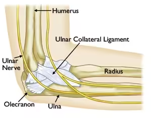
How do UCL injuries occur?
UCL injuries are very common for throwing athletes and those that use their upper extremity frequently. They can result from an acute injury (sudden onset) or from a chronic injury (over time). An acute injury can occur if too much stress is put on the elbow in one single incident which can cause the ligament to tear. An audible ‘pop’ typically can be heard or felt with this type of injury and intense pain may accompany this.
A chronic injury typically occurs from repeated stress on the ligament from throwing or swinging the arm over time. This causes the ligament to be stretched, frayed or torn. If the ligament is stretched, the repetitive motion of throwing can cause small micro tears in the ligament which can result in rupture of the ligament. Chronic injuries are more common than acute injuries for this ligament.
If there is an injury to the UCL it is often classified as a sprain. There are three grades of sprain: grade 1, 2 and 3. A grade 1 sprain means that the ligament is stretched but no tear is felt. A grade 2 sprain indicates the ligament is stretched and a partial tear could be present. A grade 3 sprain indicates there is a complete tear of the ligament.
What are the symptoms?
The most common symptom of a UCL injury is pain on the inside (medial) part of the elbow, especially during the acceleration phase of throwing. Swelling can also be present. If an acute injury occurred, swelling and bruising may also be noticed. A sensation of popping, grinding or clicking can sometimes be felt when throwing.
How are UCL injuries diagnosed?
If you notice any of the symptoms mentioned above, seeking an evaluation by a physician is the next step in diagnosing the injury. The physician will most likely do x-ray’s to rule out any type of fracture, stress reaction or loose fragments in the joint. During the physical exam portion, the physician will do several special tests to evaluate for a UCL injury. The most common test is called the Valgus stress test. A force is placed on the outside of the elbow which “opens up” the inside part of the elbow. If the UCL is working properly, it should stop the joint from opening up. If laxity (looseness) is felt during this test, it is indicative of a UCL sprain or tear. If laxity or pain are felt, this is indicative of a UCL sprain or tear. There are other special tests that can be performed, but the Valgus stress test is the most common.
If there is pain along the inside of the elbow and the Valgus stress test is positive, the physician will typically order an MR arthrogram. This is an MRI but dye is added through an injection into the elbow joint. The dye helps to make the test more accurate in that it goes into the space where a tear could be to make the tear easier to detect. If there is a tear present, surgery may be the best option to repair the tear.
How are UCL injuries treated?
UCL sprains can be treated conservatively or surgically. Conservative treatment includes rest, anti-inflammatories, physical therapy and bracing. Initially, rest and anti-inflammatories can be used to help reduce the pain and swelling. If the injury is acute, a sling may also be useful to aid in resting the elbow. Physical Therapy is a great resource for UCL injuries because it helps strengthen the muscles surrounding the elbow. This makes the elbow stronger and in turn helps to decrease the chance for further injury. A Physical Therapist can also evaluate the throwing mechanics to see if any changes need to be made in order to reduce the amount of stress on the elbow. Bracing can also be considered for support of the elbow or to reduce the amount of motion the elbow can have.
If there is a complete tear of the ligament or if pain and dysfunction persists despite conservative treatment, surgical intervention may be considered. This would include repairing the tear or reconstructing the tear. Many people are familiar with the name of this surgery which is called Tommy John’s surgery. The name is coined from the first person to ever have this type of surgery. This repair is performed when the ligament has completely torn off the bone. This is done by making an incision on the inside part of the elbow and reattaching the ligament to the bone. Sutures are used to keep the ligament in place when reattached to the bone. A reconstruction is performed when the ligament is unable to be reattached to the bone. A ligament in your wrist, called the Palmaris Longus, is often used to replace the UCL ligament if a reconstruction is performed.
What is the recovery after surgery?
Most patients are typically put in an elbow brace that limits range of motion after surgery. Each physician has different parameters for working on range of motion after surgery, but Physical Therapy will be required. The focus of Physical Therapy will be to get the proper range of motion back after surgery, although some patients may notice they have less range of motion than before. After this is achieved, the main focus will be strengthening the muscles surrounding the elbow and getting the person back to functional activity.
What’s the take away message?
Injuries to the UCL of the elbow are common, especially among throwing athletes and those who have to use their upper extremities for their sporting activity. If you notice pain along the inside of your elbow, you should seek treatment from an Athletic Trainer, Physician Assistant, Primary Care Physician or Orthopedic Surgeon. These healthcare providers all can evaluate the injury and formulate a treatment plan to help you get back to your sport.
Spring Sports Introduce New Ankle Injuries
March 21, 2017
What you need to know


Sun and warmth are just around the corner and that means high school and club sports are gearing up. Because these sports are outdoors on a field and often uneven terrain, the ankle is particularly susceptible to injury.
“Basketball and football are the sports we think of commonly to sprain an ankle, but with year round play and early spring competition we are seeing these injuries more consistently,” says Adam G. Miller, M.D., Orthopaedic Surgeon and Foot and Ankle Specialist with Beacon Orthopaedics and Sports Medicine.
Baseball, soccer, track are often played on fields that have undulations after the winter. These unsuspecting “landmines” can lead to a serious injury.
And sometimes serious injuries can go unnoticed. Miller cautions: “A rolled ankle may be the tip of the iceberg for the patient. It’s important to assess the entire lower extremity to ensure this doesn’t become a chronic issue.”
Here are some tips for ankle injuries:
- Initial protection is needed. Immediately after the ankle injury, apply ice and compression to the leg pain. Protection and rest is advisable until one knows the severity of the injury.
- Need to walk before you can run. After an injury if you can’t put weight on the leg to walk, it is advisable to make an appointment with a medical professional. This can be a sign of a complete ligament tear or subtle fracture.
- Beware of the plateau. If you are successfully improving following an ankle injury but then notice your recovery stops, don’t settle for partial relief. Chronic pain can be a sign of worsening injury such as a high ankle sprain, peroneal tendon tear or cartilage damage with osteochondral defect and should be evaluated.
- Learn from the past. While each new injury can recover similarly to previous ankle sprains, a history of multiple injuries can predispose a patient to more pain and instability that can be difficult to self-treat. A long history of ankle issues should trigger an office visit.
Bottom line: beware of the unsuspecting ankle injury that masquerades as a minor ankle “roll” and seek medical attention promptly.
For more information or a personal evaluation, please visit our website. Appointments can be made with Dr. Adam G. Miller by calling (513)-354-3700 or booking online here. If you are not following Dr. Adam G. Miller on social media, you can do so on Facebook or Twitter for: updates and comments on cutting edge treatments, discussion of various injuries, and sport/athlete issues.
Shoulder Labral Tears
March 21, 2017
Shoulder labral tears are a common acute injury. Learn more about how tears occur and some of the best options to fix them.
What is the Labrum?
Let’s start simple: The shoulder joint is comprised of 3 major bones: wing bone (scapula), collarbone (clavicle) and upper arm bone (Humerus). The Labrum surrounds the shoulder socket (Glenoid). The Glenoid Labrum is a bumper of cartilage that surrounds the shoulder joint and is pear-shaped. The purpose of the Labrum is to help stabilize the shoulder joint and keep the Humerus in the socket.

How do tears occur?
Labral tears can occur from an acute injury (sudden onset) or chronic injury (occurs over time). Acute injuries commonly occur from a fall on the shoulder when the arm is outstretched, lifting something heavy, or taking a blow to the shoulder. Chronic injuries often develop in a throwing athlete or a person that does repetitive overhead motion for their job. Oftentimes when a person dislocates (pops out of joint) their shoulder, a Labral tear results from the injury.
What are the types of tears?
There are several different types of Labral tears that can occur. These include SLAP tears, anterior tears, posterior tears and Bankart tears. SLAP tear stands for “Superior Labrum Anterior Posterior” which means that the tear is in the upper part of the labrum and extends to the back and the front of the Labrum. An anterior tear means that the front part of the Labrum is torn and a posterior tear means that the back portion of the Labrum is torn. Bankart tears usually result when a person dislocates their shoulder.
What are the symptoms?
Most common symptoms of a labral tear include a pain/ache that’s deep inside the shoulder joint, loss of velocity when throwing, and instability of the shoulder (feeling that the shoulder is going to ‘pop out’). Other symptoms include a popping or clicking sensation inside the shoulder, pain with overhead activity, pain in the front or back of the shoulder depending on where the potential tear is, and trouble sleeping at night.
How is a tear diagnosed?
The best way to diagnose a labral tear is seeking an evaluation from an Orthopedic Physician. The Orthopedic Physician will perform a clinical exam to evaluate for a tear. In order to do an evaluation, the Physician will perform several common orthopedic special tests that indicate a labral tear could be present. These common tests include Obrien’s test, Jerk test and Mayo test. Obrien’s test is performed in order to rule out a tear in the front part of the shoulder. Jerk test is performed to evaluate for instability in the back portion of the Labrum. The Mayo test is performed in order to evaluate for a Labral tear in the front of the shoulder as well.
If the physician suspects a labral tear, further imaging is needed to confirm the diagnosis. Further imaging in the form of an MR arthrogram is typically ordered. The MR arthrogram is a little different than a typical MRI because dye is injected into the affected joint. This dye adds to the imaging test because the fluid from the dye can sneak under the tear and make it easier to detect on the images. This is the best way to interpret a labral tear short of having surgery.
How is a labral tear treated?
Labral tears can be treated conservatively or surgically. Conservative treatment includes anti-inflammatory medication, physical therapy, steroid injections and rest. If conservative treatment doesn’t reduce the patient’s pain, surgical intervention may be necessary.
Surgical intervention means that an arthroscopic surgery will be performed to repair the labral tear. Anchors are used so that sutures can be passed through them in order to tie the torn Labrum back into the socket. The amount of sutures used depends on the size of the tear. If the tear is in the Bicep region, a procedure called an Open Subpectoral Bicep Tenodesis can be performed. This involves moving the Bicep Tendon to underneath a chest muscle so that the Bicep Tendon is no longer pulling on the front part of the Labrum.
Recovery from surgery:
Recovery from Labrum Repair surgery can take up to 6 months depending on the extent of the repair. Initially after surgery, the patient will be in a sling for 6 weeks. After that, the patient will have a lifting restriction of 5 pounds for the next 6 weeks and then a 10 pound lifting restriction for the 3 months following. At 6 months from surgery, the patient will most likely be cleared for all activity with the understanding that it may take up to a full year from surgery for the patient to feel fully recovered. Physical therapy is also required in order to regain the range of motion and strength that’s required for a full recovery.
When should you consider surgery?
A Labral Tear can only be “fixed” by having it arthroscopically repaired. The Labrum can also be a pain generator, therefore if Physical Therapy and Steroid injections can reduce the amount of pain, surgery may not be needed. If the pain is affecting activities of daily living and/or the shoulder joint is unstable, repairing the tear may be the best option. Talking with an Orthopedic Surgeon like Dr. Robert Rolf is the best way to determine the most beneficial treatment method.
Meniscus Tears in the Knee
March 17, 2017
What is a Meniscus Tear?
The meniscus is a cartilage cushion between the femur (thigh bone) and tibia (large lower leg bone). There are two menisci in each knee. The lateral (outside) meniscus is small c-shaped cushion, whereas the medial (inside) meniscus is a larger c-shaped cushion. Both menisci have poor blood supply and will not heal if they tear due to injury or degenerative causes. A meniscus tear typically occur when the foot is in contact with the ground and there is a twisting motion in the knee. Injury may also occur if the knee is forced to bend excessively (hyperflexion). The initial injury may or may not have associated pain; swelling typically does not occur until 48 hours after accident, and may not occur at all. Pain is generated after a tear when a piece of the meniscus folds on itself, causing catching, clicking, or popping in the knee joint. Most patients have difficulty going up and down stairs, and some may not be able to fully straighten or bend the knee.

www.tornmeniscusmd.com
How are Meniscus Tears Diagnosed?
Many meniscus tears can be diagnosed clinically based on history and physical exam. Special tests that are used for diagnosing meniscus injuries include McMurray Test, Bounce-Home Test, and Thessaly Test. A physician may order x-rays and/or MRI to help diagnose, or verify clinical suspicion of, a meniscus tear. X-rays may show decreased joint space between the femur and tibia. MRIs will show the location and type of meniscus tear and help the physician decide if the tear should be removed or repaired surgically. The MRI can also help with the decision to choose conservative treatment verses aggressive surgical intervention.
What is Conservative Treatment of Meniscus Tears?
Conservative treatment typically includes anti-inflammatory medication, physical therapy, and possibly a steroid injection in the knee joint (intra-articular corticosteroid injection). Conservative treatment will not repair a tear; this option manages symptoms.
What is Meniscus Surgery?
The majority of meniscus surgeries are menisectomies. A menisectomy is an arthroscopic procedure that allows the surgeon to identify the tear and trim the meniscus until it meets the tear, thus creating a new meniscus edge. Depending on the extent of tear and location of tear, the meniscus may also be repaired. This procedure can also be done arthroscopically, as the surgeon will use sutures to repair the tear. There is a risk that the meniscus may not heal due to limited blood supply.


www.drugs.com
Why Should I Consider Meniscus Surgery verses Conservative Treatment?
Meniscus tears will not heal on their own. Conservative treatment is beneficial to patients that do not have a decrease in motion in their knee. Anti-inflammatories can help control the pain that may be associated with the meniscus tear. Surgical treatment will remove or repair the tear.
What are my restrictions after a Meniscus Surgery?
Removal of the meniscus tear leads to limited healing time. Most people can be active in their normal routine within 6-8 weeks. Physical therapy will start almost immediately following surgery, and crutches can be used for up to a week. Repair of a meniscus tear has a longer return to activities of daily living. There is a risk of tearing the sutures used to repair the meniscus, therefore the patient is usually in a straight leg brace and non-weight bearing using crutches for at least 3 weeks. Hyperflexion or hyperextension (straight leg) can cause the sutures to catch on the femur and possibly ruin the repair. Recovery after a meniscus repair may take 3-6 months. Physical therapy for both surgical options involves strengthening of the quadriceps and hamstring muscles
What are the risks of Meniscus Surgery?
Complications are rare, but the potential does exist. Common complications may include: infection, decrease range of motion in the knee, rupture of repair, and swelling. Benefits of meniscus surgery commonly outweigh the risks.
Written By: Lisa Osterbrock, PA-C, ATC 9/18/14
Edited By: Robert Rolf, M.D.
What is a Biceps Tendon Rupture and How is it Fixed?
March 17, 2017
The biceps muscle group (biceps brachii) is made up of two heads, the long head and the short head. The muscles originate at the shoulder and insert on the radius (short bone in the forearm). The main function of the biceps brachii is to supinate the forearm (pretend like you are holding a cup of soup in the palm of your hand; this position is called “supination”). The second function of the biceps brachii is to flex (bend) the elbow. Rupture of the biceps tendon can occur proximally (at the shoulder) or distally (at the elbow). Ruptures commonly occur when there is an unexpected force applied to the bicep muscle such as attempting to catch something or someone when they fall. Most ruptures occur when the elbow is in a flexed position. Some people may feel or hear a “pop” when the tendon separates from the bone.


How is a Biceps Tendon Rupture diagnosed?
Ruptures of the biceps tendon can usually be diagnosed through history and physical exam. Observation of the injured extremity may reveal significant bruising and swelling, as well as physical deformity. The physical deformity associated with biceps ruptures is termed popeye deformity. The deformity is due to the tendon retracting toward the muscle belly causing a large bulge. X-rays may be ordered to rule out an associated avulsion fracture. An MRI may also be ordered to aid in visualizing the amount of damage sustained to the muscle, tendon, and bone.


Image courtesy of mdguidelines.com Image courtesy of eorthopod.com
What is Biceps Tendon Repair surgery?
Distal biceps tendon ruptures typically require surgical fixation to restore range of motion and strength to the elbow. This procedure is an open surgical procedure which can be performed on an outpatient basis. The goal is to reattach the tendon to the radius bone using either sutures or anchor with sutures.


Proximal biceps tendon ruptures can be treated conservatively with physical therapy and anti-inflammatories. Surgery is considered when a patient continues to have pain despite conservative measures. The biceps tendon is not reattached to its original origin; the biceps is attached to the humerus in a procedure called a “biceps tenodesis.”
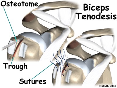
Why should I consider Bicep Tendon Surgery?
Distal repair for a biceps tendon rupture should be considered in patients that injure the bicep in their dominant arm, or active individuals. The goal of surgery is to restore strength and endurance to the musculature as well as maintain range of motion in the elbow joint. Without surgery, there is a significant probability that function of the injured extremity will be limited. Proximal bicep tendon repair is considered when conservative measures fail or do not relieve pain.
What are my restrictions after a Bicep Tendon Surgery?
Total healing time for any injury or surgical repair is usually one year. Limitations vary based on distal or proximal repair. Both repairs for a biceps tendon rupture will require the use of a post-operative sling for up to 4 weeks. Physical therapy will begin range of motion and strengthening activities based on protocols established by Dr. Rolf, or your preferred surgeon.
What will be my recovery time?
Distal and proximal bicep tendon repairs are performed as an outpatient surgery. Formal physical therapy can begin within 2 days of surgery. Formal therapy can last 12-16 weeks with gradual progression to a home exercise program. Most patients are able to return to normal activity, without restriction, 14-20 weeks after surgery. The variable wide range of return to activity is based on a distal or proximal bicep repair.
What are the risks of surgery to repair a biceps tendon rupture?
Complications associated with bicep tendon repair are rare. Common risks associated with a distal repair include: decreased strength, decreased nerve sensation, and decrease range of motion at the elbow. Common risks associated with proximal repair include: decreased strength, decreased nerved sensation, and decreased range of motion at the shoulder. Both surgical techniques have associated risk of pain, infection, and slow wound healing.


Written By: Lisa Osterbrock, PA-C, ATC
Edited By: Robert Rolf, M.D.
Images courtesy of proactivept.com, eorthopod.com, and mdguidelines.com.
Shoulder Impingement
March 15, 2017
What is a Shoulder Impingement?
Shoulder impingement occurs when the tunnel between acromion process off of the shoulder blade and the humeral head is small or the structures that run through the tunnel are inflamed. The tunnel is known as the subacrimial space. The structures that become inflamed are the subacromial bursa, supraspinatus muscle, and/or biceps tendon (long head). The subacromial bursa is a fluid filled sac that prevents wear and tear of muscle tendons as they move around a bone. The supraspinatus muscle is the main rotator cuff muscle that originates on the top of the scapula (shoulder blade), runs under the clavicle (collar bone), and inserts on the humeral head. The biceps tendon can pinch between the clavicle and humeral head. Impingement injuries commonly occur in patients that participate in overhead activities (working on ceiling fixtures, throwing motions). Overuse leads to inflammation causing a pinching pain when the arm is lifted above the head or brought across the front of the body.

http://morphopedics.wikidot.com
How is Shoulder Impingement diagnosed?
Shoulder impingement is a clinical diagnosis based on history and physical exam. Special tests that are used for diagnosing shoulder impingement include Neer’s, Hawkins-Kennedy, and Speed’s tests. Diagnostic testing may include x-rays or MRI to help rule out involvement of surrounding structures or other injury.
How is Shoulder Impingement treated?
Conservative treatment typically includes anti-inflammatory medication, physical therapy, and possibly a steroid injection (intra-articular corticosteroid injection) in the subacromial joint space. Range of motion is limited so that movement of the shoulder is pain free. The patient may be limited, not permitting the patient to lift their arm above shoulder level. Lifting the arm above shoulder level pinches the anatomic structures leading to inflammation preventing resolution of impingement.

Is surgery required for Impingement?
Surgery is not required for impingement. However, if conservative treatment fails, and the patient is still having significant pain, elective surgery may be the next option. Surgical options include a distal clavicle resection, subacromial decompression, and/or bursectomy, all of which are arthroscopic procedures. Distal clavicle resection removes a piece of bone from the clavicle; this allows an increase in size of the space below the clavicle decreasing pressure on the underlying anatomy. Subacromial decompression removes the undersurface bone from the acromion, which may include bone spurs (increased calcium formation). This procedure opens the subacromial space allowing the biceps, supraspinatus, and bursea to move freely without friction, thus preventing inflammation. A bursectomy is the removal of a bursea sac; for impingement, the subacromial bursea is removed.

www.methodistorthpedics.com
What are my restrictions after Impingement Surgery?
Patients will be immobilized with the use of a post-operative sling immediately following surgery. The sling is provided to decrease stress on the shoulder while healing occurs, as well as aid in decreasing soreness. Physical therapy will start within the first week following surgery, beginning with simple range of motion exercises and basic strengthening techniques. A patient can expect to be removed from the sling within 4 weeks of surgery, and have normal range of motion within 3 months. Pain is the most limiting factor following surgery for impingement
What are the risks of Impingement Surgery?
All surgeries have associated risks. Luckily, impingement surgery has few complications and the benefits typically outweigh the risks. Common risks and complications include bleeding, infection, swelling in the elbow/wrist/hand, and numbness/tingling in the elbow/wrist/hand.
See if you’re dealing with shoulder impingement and what the next steps are, by scheduling an appointment with Dr. Robert Rolf.
Common Questions About MCL Knee Sprains
March 15, 2017
What is a MCL Sprain?

MCL stands for medial collateral ligament and is located on the inside of the knee. The MCL originates on the femur (thigh bone) and inserts on the tibia (large bone of the lower leg). The MCL splits slightly at the knee joint and some fibers also insert onto the medial meniscus (cushion inside the knee). Due to attachment of the MCL on the medial meniscus, MCL sprains may also have an associated medial meniscus tear. An MCL sprain occurs when there is a direct force applied to the outside of the knee, pushing the knee inward. MCL sprains may also occur when a person sustains an ACL (anterior cruciate ligament) tear. There are 3 grades of sprains. A grade one sprain involves stretching and minor tearing of the MCL fibers; a grade two sprain involves partial (50%) tearing; and, a grade three sprain is a complete tear or rupture of the MCL. After sustaining any type of MCL injury, there may be difficulty bending and straightening the knee.
How are MCL Sprains diagnosed?
MCL sprains can usually be diagnosed clinically via physical exam. The health care provider will perform a special test called a valgus stress test. This test may reveal pain and/or instability on the medial side of the knee. X-rays may be ordered which may reveal an avulsion fracture; this involves the MCL pulling a small chip of bone off of the femur. A MRI may be ordered to verify the extent of tearing that has a occurred to the MCL and will also diagnose any additional associated injuries.
How is an MCL Sprain treated?
Most MCL sprains will heal with conservative treatment. These sprains include grade one and grade two sprains. Conservative treatment involves non-weight bearing with the use of crutches until the patient is able to walk without a limp or pain, and physical therapy to strengthen the surrounding musculature (quadriceps, hamstrings, adductors (groin)). The patient may also be placed in a hinged knee brace to help protect the knee from side to side motions. A grade three sprain/tear may require surgical repair.
Why should I consider MCL Surgery?
If someone has a complete tear of the MCL conservative treatment may be an option. The leg would have to be placed in a knee immobilizer, typically locked in full extension, for several weeks to allow scar tissue to form and repair the MCL. After scar tissue has formed, the patient will need to attend physical therapy to strengthen the surrounding musculature. Surgical treatment involves repair of the MCL or reattachment of the ligament to the bone. Surgical repair is not done arthroscopically; it is an open procedure. Surgical repair is preferred for athletes that require excessive amounts of twisting and pivoting at the knee joint.
What are MCL surgery post-surgical restrictions and recovery?
Following MCL surgery, the patient will be placed in a knee immobilizer that is locked in slight flexion (bent knee). The brace can be unlocked at the discretion of the doctor and physical therapist. With the brace locked in flexion, the patient will be non-weight bearing with the use of crutches for 4-6 weeks. The brace can be slowly unlocked to increase range of motion at the knee at the doctor or therapist’s discretion. Once the patient is permitted to weight-bear, strengthening, range of motion, and balance, are key areas that are addressed in therapy. Aggressive therapy usually does not occur until 12 weeks after surgery. It is important to realize that it may take up to a year for a patient to have full recovery following surgical repair of the MCL.
What are the risks of MCL Surgery?
Complications or risks are associated with any surgery. Common risks include: infection, loss of range of motion, and instability. In most cases, the benefits of surgery out weigh the risks.
Written by: Lisa Osterbrock, PA-C, ATC 9/23/14
Edited by: Robert Rolf, M.D.
The Advantages of an Arthroscopic Procedure
March 14, 2017
For high school and college athletes, there are few things worse than a season-ending injury. If sitting on the sidelines wasn’t already painful in its own sense, a severe injury and the conditions associated with it may continue long after the season has ended and may even lead to problems as an adult.
Orthopedic injuries are an unfortunate reality for the millions of individuals who participate in professional sports or even just recreational activities. While surgery is not necessary for the vast majority of injuries, a diagnosis by a board certified sports medicine physician can determine if and what type of surgery may benefit you in the present as well as prevent more serious problems down the road. Dr. Steve Hamilton is a fellowship trained sports medicine physician at Beacon Orthopaedics and Sports Medicine who can determine if your injury requires surgery. In most instances, Dr. Hamilton will recommend arthroscopic surgery over an open procedure.
Arthroscopy, which is also called “arthroscopic surgery,” is one option that you can discuss with your sports medicine physician. This minimally invasive surgical procedure is often used to examine and treat a patient’s shoulder, hip, wrist, or ankle; however, it can be performed on most joints.
Arthroscopic surgery can treat but is not limited to:
- Damaged anterior cruciate ligament (ACL)
- Damaged medial collateral ligament (MCL)
- Damaged ulnar collateral ligament (UCL)
- Torn or damaged meniscus
- Irritated, enlarged, or inflamed knee (Plica syndrome)
- New cartilage stimulation (Microfracture)
- Cartilage transfer (Autologous chondrocyte implantation)
- Loose cartilage
- Damaged or deteriorating cartilage
- Torn rotator cuff or labrum
- Arthritis
- Tendonitis
- Bursitis
- Bone fractures
- Bone spurs
- Baker’s cysts
- Ganglion cysts
Arthroscopy is considered a minimally invasive procedure because it allows the surgeon to both view and treat a joint without fully opening it up. This means less trauma to the joint and less scarring, and often provides shorter recovery times and lower risks of complications compared to open surgeries.
How Arthroscopy is Performed
Pre-Procedure
If arthroscopy is the best method of treatment for you, you will be given general anesthesia prior to the procedure. Your orthopedic surgeon or anesthesiologist will talk to you about which option is best for you.
The surgery center at Beacon Orthopaedics provides viewing rooms behind glass for family members to view your procedure. The rooms are staffed with surgical nurses who are able to answer any questions as they walk the family through the procedure.
Procedure
Once the patient has been properly draped and the skin around the joint has been thoroughly cleaned, the orthopedic surgeon making a small incision. These incisions are significantly smaller than the ones made during open joint surgery. At this time, the surgeon will also fill the joint with irrigation fluid which will distend the joint and rinse away any cloudy fluid obstructing their view.
Next, the surgeon inserts an arthroscope, which has a diameter that is approximately the same size as a pencil’s, into the opening. This instrument contains lighting and a camera, transmitting video of the interior structures to video monitors so the surgery team can see inside the joint. It also contains a small lens and a light which allows the surgeon to magnify and illuminate difficult areas.
In some instances, the surgeon may also need to remove a small amount of tissue in order to view or access the joint. In order to do this, they use a specialized shaver to resect, or remove, the smallest amount of tissue that’s required.
This allows the surgeon to a full view of the joint for a procedure or debridement (removal of damaged tissue and debris) with a very minimum incision.
Closure
In general, the amount of time it takes to perform arthroscopic surgery is significantly less than that of traditional joint surgery; however, this will depend on any unexpected findings and what type of treatment is performed.
Once your joint has been successfully restored or repaired, Dr. Hamilton will close your incision and cover it with a soft bandage. Most patients are safe to remove the bandage the following morning.
The Benefits of Arthroscopy
Lower Risk of Complications
The smaller incisions made during an arthroscopic procedure mean that there is a lower chance of an infection. Moreover, the procedure is simpler and more straightforward than a traditional approach. This is primarily due to the fact that no muscles or tendons need to be cut.
Faster Recovery Time
Your incisions will heal within a day or two of your operation and you will likely be able to return to your daily activities shortly afterward. Arthroscopy is almost always performed as an outpatient procedure. This means you can go home the same day as your operation. Dr. Hamilton may prescribe physical therapy to help expedite your recovery. If you are an athlete, a more rigorous rehabilitation program may be designed to help you return to play. Keep in mind, however, that the actual length of time that your body will need in order to heal fully depends on the specific condition that caused your problems as well as your own unique health factors.
Less Post-Operative Pain
Alongside a faster recovery, arthroscopic surgery often results in less pain during the first few days following the procedure. While you should still expect some pain and swelling, these symptoms are noticeably easier to manage, especially compared to the post-operative pain that is commonly experienced after open surgery. This is primarily due to the fact that arthroscopic procedures are less invasive and less traumatic than open surgeries.
Candidates for Arthroscopic Surgery
Candidates for arthroscopic surgery share many qualities with candidates of other minimally invasive procedures, such as platelet injections treatments. These candidates tend to be younger, more active, and generally healthy. These qualities are ideal for a few reasons: First, the surgeon must be able to navigate the arthroscope and other necessary tools through the interior structures of the patient’s body. Second, physical therapy is a common part of the patient’s rehabilitation. A higher level of movement before surgery usually results in a faster recovery.
Talk to Dr. Hamilton about Your Arthroscopic Options
While all forms of surgery carry a level of risk, an arthroscopic procedure conducted by an experienced, fellowship trained surgeon can be a highly effective way of treating joint problems.
Dr. Steve Hamilton is not only one of the most recognized arthroscopic surgeons in the Greater Cincinnati and Northern Kentucky area, but he also joined Beacon Orthopaedics and Sports Medicine as the first sports medicine physician in the area with fellowship training in hip arthroscopy. He can discuss with you whether minimally invasive joint surgery is the best option and provide specific expertise regarding shoulder, hip, knee and ankle injuries.
Learn more about Dr. Hamilton or schedule an appointment to meet with him at Beacon East, Beacon West, or Summit Woods in Ohio or at Beacon’s Northern Kentucky location.
Back Pain at Work: 3 Ways To Reduce It
March 8, 2017
 You were designed to be active. Unfortunately, despite the substantial body of research which concludes that a sedentary lifestyle leads to severe back problems as well as other serious health conditions, the average person spends approximately 55% of their waking hours sitting, according to a study by Vanderbilt University. Maintaining a static position for too long causes stiffness and tension to develop around the spine and can consequently lead to a short-term, or even a permanent, lower back condition.
If a sedentary lifestyle is the cause of lower back pain, why do so many of us spend our waking hours sitting? To understand why, look no further than modern work habits. Many of us spend 8 hours a day – if not more – sitting at a desk as part of a typical work or school day. And while there is nothing inherently wrong about using an office desk and computer to conduct business, how our working area is set up, our posture, and the level of activity we engage in throughout the day all put our spine at risk.
Fortunately, these are all factors that we can control.
1. Adjust Your Workspace
Proper care for your back starts with adjusting your work environment. Whether you use an office desk or another type of workstation, you need to set it to a height that you will be most comfortable working at for long periods of time.
Once you have determined the optimal height of your desk, you will then need to select appropriate seating. Ergonomic chairs are an excellent choice; however, keep in mind that simply owning one is not enough to preserve your back. Whether you use a traditional office chair or an ergonomic option such as a Swedish kneeling chair or a Swiss exercise ball, you will also need to adjust it to your individual proportions.
Here is how you can ensure that your chair is properly adjusted.
Adjust the height of your chair
Sit as close to your desk as you comfortably can. With your upper arms parallel to your spine, place your hands on the surface of your desk. Your elbows should form a 90-degree angle. If your elbows hang lower than the surface of your desk, adjust the height of your chair either up or down until your arms form the correct angle.
Adjust your armrests
An ideal chair has armrests and they should be adjusted to slightly lift your shoulders. Doing so will not only reduce the strain placed on your upper spine and shoulders, but it will also make you less likely to slouch forward.
Adjust your backrest
With your body situated against your backrest, form a fist and place it between the back of your calf and the front of your chair. If you do not have enough space for your fist, your chair is too deep and the backrest needs to be adjusted forwarded. If the back of your chair cannot be adjusted, you can use a cushion or a rolled up towel to achieve low back support.
Determine your resting eye level
While you are still situated in your chair, close your eyes for a few moments and then slowly open them. As you open your eyes, your gaze should be on your computer screen or the area where you will be looking for the majority of your work. If this area is not aligned with your resting eye level, or if you sense that you are tilting your head either higher or lower, you will need to make adjustments accordingly.
Position objects within arm’s reach
By keeping the items you use most frequently within arm’s reach, you can reduce your need to stretch and consequently reduce your risk of straining a ligament. At the same time, this prevents the need to “break” your current position and thereby helps you maintain correct posture for longer periods of time throughout the day.
2. Practice Correct Posture
While the optimal setup of your office chair and desk are important, preserving the health of your spine also requires active muscle use in order to maintain correct posture. Slouching forward or slumping back strains the muscles, ligaments, discs and other components of your back. Over time, poor posture while sitting can even increase the amount of stress placed on other areas of your body, including your shoulders, arms and legs.
To minimize the amount of stress placed on your back, you should sit as close as possible to your desk with your head upright, your upper arms parallel to your spine, and your hands rested at a 90-degree angle on your work surface. Your legs should also be positioned at 90 degrees, directly above your ankles. If you properly adjusted your workspace in accordance with the aforementioned guidelines, you will already be in the correct position.
3. Stay Active
While correct posture will minimize the amount of gravitational force placed on your spine, it will not solve your problem of simply being stationary for too long. While you should take every opportunity to be active at work, at a minimum you should stretch at least once during the first half of your day, once in the middle, and once towards the end. If you find it difficult to set aside a few minutes in your day to stretch all of the areas of your body, you can stretch one area at a time as you transition from one room or another or as a dedicated portion of your lunch.
While stretching will help relieve your body of the aches and pains that develop from being stationary, work-related back pain can also be the result of stress. Between tight deadlines, budget demands, performance reviews, or even just the ordinary challenges of the day, it’s easy for stress to build up in the body. You can combine breathing exercises and other relaxation techniques with your stretches to keep both your mind and, by extension, your body healthy.
Conclusion
Our time at our job or at school comprise the majority of the time we spend sitting. By adjusting your work environment and your personal habits, you can significantly reduce the amount of stress placed on your back over your lifetime. Keep in mind, however, that excessive sitting, whether at work or during your leisure, is harmful. Because of this, it is also important make the necessary adjustments in other areas of your life in order to ensure complete care for your back.
If you require more specialized spine care, we can guide you towards an appropriate treatment. As a board certified orthopedic surgeon for over a decade with a sub-specialty in Spinal Surgery and Management of Spinal Conditions, wee can diagnose the cause of your back problems and effectively treat your condition. While a very small amount of cases actually require surgery, we will be able to help you determine the best course of action for your particular situation.
Non-Surgical Treatments for Back Pain
March 3, 2017
Your back works hard. In fact, your lower back carries the entire weight of your torso throughout the day, including the time you spend sitting, standing and moving. Unfortunately, our everyday activities as well as the amount of time we spend sitting at work, during our commute and during our leisure make it easy for us to place excess stress on our spine. Sometimes all it takes is just a little bit too much stress in order to cause mild, temporary back discomfort or even a constant, excruciating pain that may develop into chronic back pain.
With all of this in mind, it may be unsurprising that experts estimate that as much as 80% of the population will experience a back problem at some point in their lives.
At Beacon Orthopaedics and Sports Medicine, workplace injuries are one of the primary reasons that patients come to us for back and spine care; however, we also recognize that poor posture, improper body mechanics, or a combination of other factors can all be reasons why individuals throughout Greater Cincinnati turn to us for treatment. In the vast majority of cases, back problems can be treated without the need for surgery.
Before starting any kind of treatment plan, it’s important to make an appointment with one of our board-certified, sub-specialty trained physicians. The following information is intended to help guide your discussion.
Home Remedies for Back Pain
Approximately 80% of all back pain is acute which means that it results from muscle spasms or minor strains or tears in your muscles or ligaments. Acute pain, which can range from mild discomfort to debilitating pain, often comes on suddenly and can last for up to 5 weeks. In most instances, minor problems will improve within 7 days with proper rest.
Of course, there’s never a “good” time for an orthopedic problem, and while it may be comforting to know that your condition will resolve itself with time, our team at Beacon recommends that you always have a plan for orthopedic and spine care. Here are a few steps you can take as a part of your plan for acute back pain.
Apply Heat
Heat is an effective way to soothe muscles and connective tissues in the days following a back sprain or strain. Heat should be applied to the site of your discomfort for up to 20 minutes at a time throughout your day utilizing either a hot pack, heating pad, warm bath, or shower.
This type of therapy is effective because it dilates blood vessels and helps facilitate the flow of oxygen and nutrients to your muscle. Heat can also help the soft tissues around your spine become more flexible and consequently bring comfort to your back.
Stretch
Regular stretching is effective for both easing your current pain as well as preventing future injuries. Stretching will loosen tense muscles, strengthen those that need improvement, and increase your range of motion. Stretches should be done at the start of everyday physical activities, before exercise and whenever you need temporary relief.
It is important to discuss your condition with a physician or orthopedic specialist prior to beginning an exercise or stretching routine. Depending on the location and the cause of your back pain, some stretches may provide you with relief while others may actually worsen your condition. Stretches should always be gentle. If, at any time you experience pain resulting from your activity, stop immediately. A Beacon physical therapist can demonstrate proper stretching techniques to remain mobile, reduce your pain, and prevent injury.
Limit Bed Rest
When you experience pain, the natural inclination is to lie down and take it easy; however, inactivity not only undermines your body’s natural healing process, but it can also be potentially harmful. A lack of activity can reduce your muscle strength and endurance and consequently make it more difficult for your back to support you once you’re on your feet again. Moreover, excessive periods of rest can lead to degenerative joint disease, the development of blood clots, and other serious complications.
Fortunately, all of this can be avoided by remaining active and limiting bed rest. When your pain is severe, your physician may advise that you spend up a day or two resting and return to your everyday activities as soon as you are able.
Manage Your Stress
Back aches and pains aren’t exclusively caused by injury. At times, stress can be a contributing factor to our condition if not the root of it. This is due in part to stress’s ability to cause the muscles, connective tissue and adhesions around our spine to become stiff. This can aggravate nerves and restrict the flow of blood to the affected area.
Of course, this means that relaxation techniques can help you remain calm and avoid potential problems. Meditation, in particular, can also help you improve your tolerance for pain when it does arise as well as your perception of your symptoms.
If your stress stems primarily from a physically demanding job or sport, it’s important to not push yourself too hard or unnecessarily. Scheduled breaks can be an effective way of balancing an active lifestyle with the strain that may result from it.
Professional Back Pain Treatment
Unfortunately, not all back problems resolve within a few weeks. Subacute pain typically lasts 7 to 12 weeks. Chronic pain, by contrast, lasts approximately 12 weeks or longer. In either case, the degree of your pain can range from mild to severe.
These conditions are often the result of an isolated injury or a combination of factors, such as years of poor posture, being overweight, and sleeping on a poor mattress. In some cases, it can also be the result of natural, degenerative changes due to aging or an underlying disease.
If you experience a significant injury or if your acute pain does not improve after several weeks, contact a Beacon Orthopedics spine physician immediately. Here are two of the non-surgical treatments for back pain that Beacon can provide.
Physical Therapy
The goal of Beacon’s physical therapy program is to relieve your pain, increase your ability to function, and educate you on techniques to prevent further injury.
For the vast majority of our patients, exercise has proven itself to be paramount to their rehabilitation. Patients who regularly undertake aerobic exercise are more likely to remain functional and also experience less pain when an incident occurs. Exercise can also improve the distribution of nutrients throughout your back as well as elevate your mood, keeping both your body and mind healthy.
The type of exercises that your treatment plan consists of will depend primarily upon your specific diagnosis and severity of pain. At Beacon, we provide our patients with the plan, guidance and the space necessary in order to exercise in a safe and progressive manner. We are also uniquely qualified to work with high school, college, and former athletes to resolve their back problems and return them to their highest level of function. In fact, our Bridge Program is designed to provide athletes with a tailored exercise plan that will allow them to seamlessly return to their activities following an orthopedic problem.
In addition to active therapy, our physical therapists also teach you correct posture and body mechanics. These techniques will help you avoid placing unnecessary levels of stress on your spine, as well as its discs, in order to preserve it and prevent future problems.
As a compliment to physical therapy, chiropractic care is offered at Beacon’s Summit and Wilmington locations. There is a substantial body of research which concludes that manual therapies are an effective way to treat lower back pain, neck pain, and other conditions. In fact, chiropractic care is highly recommended as an early treatment during the onset of your symptoms.
Injections
orthobiologics is another effective, non-surgical option for back pain that has become increasingly well known. orthobiologics entails precise, minimally invasive injections and distinguishes itself from other forms of back treatment by promoting the body’s own ability to naturally to heal its muscles, tendons, ligaments, cartilage, bones, and other tissue.
The basic principle behind orthobiologics should be familiar: When you sustain a minor cut, your cells go to work to heal your wound. When you sustain a more serious injury, a greater number of cells are involved in the healing process. By extracting the right type of cells from a highly concentrated region of the body and injecting them into the area where they will be most beneficial, your body will be more effective at naturally healing itself. Also, because ultrasound is used to locate the optimal injection site, you can be confident that the procedure is conducted with the highest level of precision and care.
A physician can identify both the source of your problem and which treatment is most appropriate for treating it.
Experience Back Improvements with Beacon
Even though we only covered a few treatments, you have many options for treating your back pain.
At Beacon Orthopaedics and Sports Medicine, our physicians, physician assistants, physical therapists, and orthopedic specialists work side-by-side in order to provide a complete continuum of care. With over two decades serving Greater Cincinnati and a specialization in orthopedic and spine care, our team can diagnose your condition, design an effective treatment plan, and guide you towards recovery. Request an appointment at any one of our convenient locations throughout Greater Cincinnati.
Our team at Beacon Orthopaedics will ensure that you experience life without back pain.
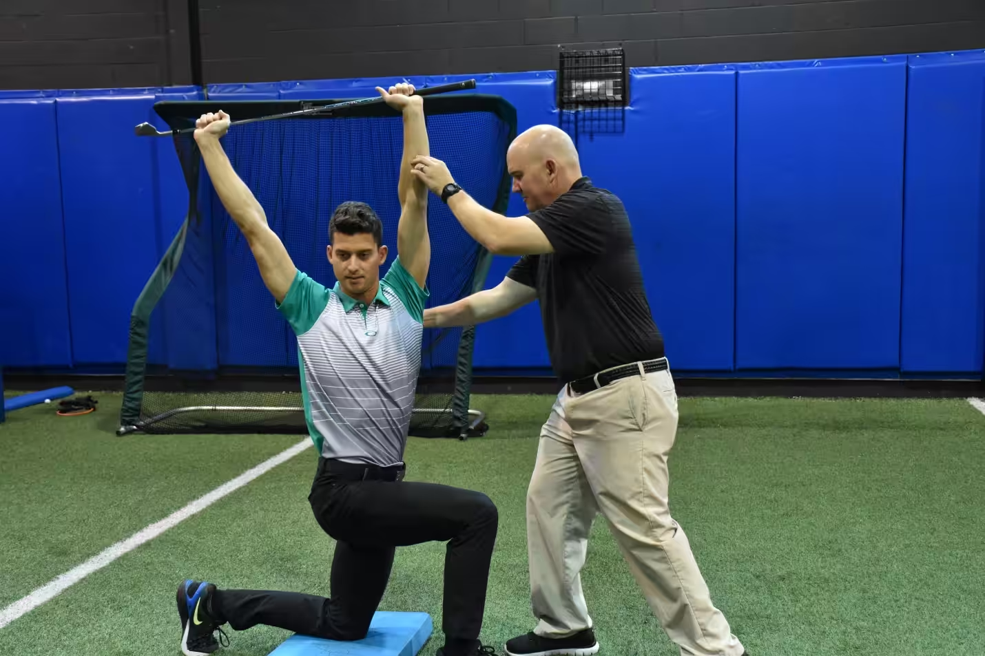
Anterior Joint Replacement: An Alternative Treatment for Hip Pain
February 22, 2017
If your hip has been irreversibly damaged by arthritis, the excruciating pain associated with movement can interfere with daily living. In fact, depending on the severity of your condition, it may impair your ability to walk, stand up, climb stairs, or complete other necessary activities. A total hip replacement, also called a total hip arthroplasty, can be a safe and effective way to return you to a normal, pain-free life when other, more conservative treatments have not helped.
At Beacon Orthopedics and Sports Medicine, Dr. Haleem Chaudhary takes a minimally invasive anterior approach to total hip replacements. This type of procedure can provide you with all of the same benefits of a traditional hip replacement, with far less tissue trauma, a faster recovery, fewer post-operative restrictions, and a lower risk of displacing your new joint. This makes an anterior approach ideal for professionals who don’t want to take too much time off of work, or active adults who don’t want to miss an entire season of their favorite sports.
While an appointment with Dr. Chaudhary is the only way to determine if an anterior approach to total hip replacement is the best option for your hip pain, here is information you can consider beforehand.
Advantages of Anterior Hip Replacement
An anterior (front) approach to total hip replacement differs from a traditional, posterior (rear) approach in a few significant ways.
During a traditional hip replacement, a surgeon would make a 10 to 12-inch incision on the side of your hip. Next, the surgeon would cut through and remove soft tissue attached to your hip bone in order to dislocate it. By doing this, the surgery team would have a clear view of your joint. Finally, the surgeon would then remove the damaged bone and insert and secure an implant in the appropriate location.
At Beacon, an anterior approach hip replacement often involves a smaller incision of only 3 to 6 inches in length. As the name implies, the incision would be made at the front of your hip area where there is substantially less soft tissue. The most significant difference with an anterior approach, however, is the fact that Dr. Chaudhary would utilize a muscle interval in order to access your joint. This would allow the surgery team to work between your soft tissue rather than cut through it.
While the limited amount of trauma done to your body is perhaps the most significant reason to consider a minimally invasive procedure, an anterior approach offers additional benefits including those following.
Faster Recovery
Naturally, less damage to your body means less time for recovery so, in addition to less potential for scarring, you can also expect a shorter stay in our overnight rooms. Nearly all patients being physical therapy the following day, and are cleared to go home within 24-hours of surgery. Ultimately, though, the amount of time you need to recover will depend on your personal health.
Better Range of Movement
Patients who undergo a traditional hip replacement often have to adhere to a strict set of instructions during the weeks following their surgery. These patients are instructed to avoid bending over, crossing their legs, or undertaking other movements that may dislocate their new joint. Additionally, a physician may instruct the patient to sleep with a pillow between their legs or use an elevated toilet seat. And while physicians often advise patients to follow these precautions for 6 to 8 weeks, some may recommend a longer period of time or require the patient to refrain from certain activities indefinitely.
An anterior approach would reduce your amount of post-operative restrictions. You could bend over, sit with your legs crossed, use normal chairs, drive, or otherwise return to everyday living within a few weeks of your operation.
Lower Risk of Hip Displacement
No matter what form of hip replacement you receive, you will need to learn how to adapt your movements in order to accommodate and preserve your implant. Fortunately, an anterior hip replacement offers a lower risk of dislocating your hip’s new ball and socket. In fact, with proper care, patients are able to preserve their new joints for years and even enjoy swimming, cycling, golfing, and other low-impact sports.
Candidates for a Minimally Invasive Anterior Hip Replacement
In order to be a suitable candidate for a minimally invasive anterior hip replacement, you should meet the criteria for surgery in general as well as meet the criteria for this specific procedure.
A good candidate for a general hip replacement has debilitating pain or restricted movement that limits or completely impairs their ability to complete ordinary tasks. This pain is most often the result of severe osteoarthritis, rheumatoid arthritis, or another form of the disease that has irreversibly damaged the joint or hip bone; however, surgery may also be an option for those with a different condition that significantly limits their movement and quality of life.
In terms of an anterior approach, a surgeon must be able to both work between soft tissues as well as operate with a restricted view of the joint area. Because of this, individuals with smaller frames are often better candidates than those with muscular or obese frames.
Keep in mind, however, that surgery is often reserved for those who have attempted other, more conservative forms of treatment such as injections, orthobiologics, or physical therapy, but experience limited or no improvements. Moreover, you will be required to complete physical therapy that will help ease your post-operative pain, restore your range of motion, and teach you how to preserve your new joint.
Meet with a Physician to Discuss Your Options
Like all forms of surgery, anterior hip replacement carries a level of risk. The only way to minimize these risks and ensure the best outcome for you is to work with a physician of considerable experience and skill.
Dr. Chaudhary has been a board certified orthopedic surgeon and a member of Beacon Orthopaedics and Sports Medicine for nearly a decade. With a specialization in minimally invasive anterior hip replacement, he can quickly determine if a joint replacement is the best treatment option for your condition. If you meet with Dr. Chaudhary, he will speak with you about what you can expect from the procedure, as well as on the day of your surgery and during the weeks following.
Learn more about Dr. Chaudhary or schedule an appointment to meet with him at Beacon East, Summit Woods, and Beacon West in Ohio or at Beacon’s Northern Kentucky location.
Non-Surgical Approaches to Arthritis
February 16, 2017
The 50 million American adults who suffer from Arthritis—no matter their background, ethnicity, or age—have one important thing in common: a desire to relieve their symptoms.
Joint replacement surgery is often considered the primary solution for alleviating the pain and lack of mobility caused by joint inflammation. If you are one of the millions of adults suffering from this condition, it’s important that you recognize that surgery is not your only option. In many cases, it doesn’t even have to be your first choice—there are many non-surgical approaches for treating arthritis.
At Beacon Orthopaedics and Sports Medicine, we have helped hundreds of patients ease the symptoms of their arthritis and preserve their joints without surgery. In many cases, changes to their lifestyle have made their condition manageable and have allowed them to return to their favorite activities. If you are considering joint replacement or another form of orthopedic surgery, here are a few alternative options to discuss with a Beacon physician first.
What is Non-Surgical Treatment for Arthritis?
Non-surgical treatment for arthritis, which is also called non-operative treatment or conservative care, includes the use of physical therapy, pain management, and orthobiologics to reduce your discomfort and increase your mobility. In contrast to surgery, this approach eliminates the potential risk, stress, and significantly higher cost of an operation. It also vastly reduces the recovery time.
The combination of physical therapy and injections will depend on the type of arthritis you have and other personal factors. However, regardless if you have osteoarthritis, rheumatoid arthritis, psoriatic arthritis, or another form of the condition, you can rest easy. There is an effective treatment plan for you.
Best of all, a non-surgical approach to managing your symptoms will provide you with many options that you can do with just an initial discussion with your physician. These include eating a healthy diet, managing your stress, and exercising regularly. For many individuals, self-management is empowering and it can give you the peace of mind that is often difficult to maintain while thinking about surgery.
Ways of Managing Your Arthritis
Meeting with a Beacon physician is the first step in your treatment plan. From there, your physician will work with you to identify which of the following orthopedic services or lifestyle changes will be beneficial to you. You will also be able to find out if you are a good candidate for non-surgical regenerative options. While changing your diet and regular exercise may not offer relief in advanced cases of osteoarthritis or psoriatic arthritis, they can provide major benefits in the early stages of chronic joint inflammation.
Eat Non-Inflammatory Foods
As many high school and college athletes understand, eating a healthy diet is vital for the overall wellness of your body. And, just as there are foods to help build your body, there are many foods that will help you maintain it, particularly your joints.
Eating a diet that is adequate in fish, nuts and seeds, fruits and veggies, olive oil, beans, and whole grains provide the immediate benefit of curbing inflammation. As a secondary benefit, a well-managed, healthy meal plan can also contribute to weight loss and consequently reduce the amount of stress you place on your joints.
Manage Arthritis Flares Due to Stress
Stress is recognized as a factor that leads to arthritis flares. Naturally, managing your stress will not only curb the pain of a flare but it may also reduce the number of times flares occur in the first place.
Unfortunately, stress manifests in our lives in a multitude of ways. An obvious form of stress arises when we take on too many responsibilities with not enough time or resources. Other, more subtle forms are the result of caffeine, alcohol, nicotine, or a lack of sleep. Whatever the source of your stress is, the first step is simply being more aware of it. From there, you can better manage it in the moment and develop strategies for managing it over the long term.
Exercise Your Joints
Joint pain and stiffness are perhaps the two most common symptoms of arthritis. One way patients seek to avoid these symptoms is through inactivity. Unfortunately, this often makes their problem even worse. While it may seem counterintuitive, exercise can be one the single most effective treatments for many forms of arthritis.
Low-impact exercises such as tai chi or yoga can help restore normal joint mobility and strength. These exercises can also provide targeted relief to your ankle, back, knee, or another area of discomfort. If you are an athlete, you will be happy to know that there are also ways to safely lift weights, run, and generally maintain an active lifestyle while managing arthritis.
The exact frequency and intensity of your workout will vary from person to person and so, similar to your diet, it is important to discuss your exercise plan with your physician prior to starting. Beacon’s specialization in sports medicines means that our doctors are uniquely qualified to answer your questions about choosing an exercise plan, proper stretching techniques, and how to avoid overtraining.
Receive Physical Therapy
In many cases, the pain of arthritis is due in part to poor movement habits. For example, a person whose heels pound the floor as they move places unnecessary stress on their knees and hips. A physical therapist can identify the harmful habits and train you to move in ways that reduce your pain and avoid additional trauma to your joints.
Physical therapy often includes manual therapy in addition to exercise and education. A certified therapist can apply pressure to your body. They will provide special exercises and stretches to help stop and, when possible, reverse joint damage and inflammation. An orthopedic specialist, in particular, is able to provide a better level of care to your neck, back, shoulders, knees, and other joints, than a general therapist.
In some cases, physical therapy has been shown to delay the need for surgery or has been proven to be just as effective as a joint replacement. A discussion with a sports medicine doctor at Beacon Orthopaedics will help you learn more about what level of physical therapy is appropriate for your arthritis.
Receive orthobiologics
orthobiologics options are non-invasive or minimally-invasive ways of healing areas of your body without the need for surgical intervention. Their benefits range from regenerating articular cartilage in arthritic joints to treating inflamed and injured nerves. This, of course, also means that this type of treatment is a viable alternative to joint replacement. Until very recently, there were no proven options for helping the body rejuvenate or grow new cartilage. These cutting edge non-surgical procedures (injections, primarily) empower the body to heal itself, instead of relying on new joints.
At Beacon Orthopaedics, we offer a number of options for orthobiologics, including:
- platelet injections therapy
- Percutaneous Needle Tenotomy (PNT)
- Prolotherapy
- Perineural injection treatment
After you have attempted more conservative methods of treating your joint pain, such as a healthy diet and adequate rest, you may consider talking to a Beacon physician about orthobiologics if you continue to suffer from joint pain, inflammation, or restricted mobility.
Specialized Orthopedic Care for Your Arthritis
Beacon physicians will work with you to develop a non-surgical approach to your arthritis. With a combination of lifestyle changes and specialized orthopedic care, you may be able to treat your symptoms without the need for open surgery.
We have 25+ locations throughout Greater Cincinnati, Dayton, Northern Kentucky, and Southeast Indiana. Request an appointment with a physician or call (513) 354-3700. Beacon Orthopaedics will help you manage your arthritis and return to an active lifestyle!

Receive Immediate Orthopedic Care at Our Walk-In Clinic
February 7, 2017
When you experience an orthopedic injury, the sooner it is addressed and the sooner the proper therapies are applied, the sooner you can recover and return to your pre-injury lifestyle. While this is especially important for college and high school athletes who want to return to their highest level of performance, it is also important for those who experience a problem resulting from the workplace or everyday activity. And while you can’t predict when you’ll need orthopedic care, you can have a plan for when you do.
At Beacon Orthopaedics and Sports Medicine, our goal is to provide timely, high-quality care for your orthopedic needs and help you return to your active lifestyle. That’s why we are proud to provide the most advanced walk-in clinic in Northern Kentucky so residents of Boone, Kenton, and Campbell counties have convenient access to orthopedic care whenever the need arises. Best of all, no appointment is required to see a specialist. Just walk in and a physician or physician assistant will start addressing your problem within minutes.
Do you have a plan for addressing an injury? Here are three reasons why a visit to Beacon’s walk-in clinic is a better choice than an emergency room visit.
Extended Hours
Whether you suffer a sudden, acute injury to your wrist during weekend golf practice or if you experience back pain from extensive hours sitting at your desk during the week, our orthopedic walk-in clinic offers extended hours that accommodate a variety of schedules. Centrally located in Erlanger, our location is also convenient to reach from anywhere in Northern Kentucky.
Whether you want care during Tuesday’s lunch break, after Thursday’s soccer practice, or on a Saturday morning, our clinic is available to you.
Instant, Specialized Treatment
 Within minutes of walking through the door, you will meet with an orthopedic specialist to address your fracture, concussion, sprain, strain, dislocation, or other orthopedic injuries.
And, unlike emergency rooms that serve a broad range of urgent health problems, our clinic is staffed by fellowship-trained physicians and licensed physician assistants. These physicians have completed years of training centered on a specific category of orthopedics, or “sub-specialty”, which allows them to provide a higher level of care than a general physician. Whether your pain is due to a sports injury, the workplace, or a chronic condition, the orthopedic specialists at our Northern Kentucky location are able to address an array of problems associated with the:
- Elbow
- Knee
- Foot and ankle
- Hand and wrist
- Hip
- Back and neck
While a trip to the emergency room is one way of addressing your problem, the long wait associated with the average visit often results in extended periods of pain, additional stress, and potential exposure to a number of viruses and pathogens. Additionally, many ER doctors will refer you to a specialist thereby delaying your treatment even further.
Cost-Savings
When you have pain, you want immediate relief. Unfortunately, a typical trip to the emergency room often results in additional pain that comes later when the bills finally arrive. While our plan for serving you includes convenient hours and the highest quality of treatment, we also do so for a cost that is far less than the cost of an emergency room visit and can accept almost all types of insurance, including Medicare and Medicaid. According to The Atlantic, the average ER visit for back problems, sprains, and strains is $1,500. At the Beacon Orthopaedics walk-in clinic, you can expect costs around 1/7 of that amount.
For us, returning you to your pre-injury lifestyle means more than just treating your pain. We want you to have peace of mind, too.
Receive Care for Your Orthopedic Injury Now
While a trip to the emergency room is one way of addressing your problem, the long wait associated with the average visit often results in extended pain, additional stress, and potential exposure to a number of viruses and pathogens. Worse, a typical visit to the emergency room can cost thousands of dollars.
And sometimes, even if you don’t have an emergency, there may be times when you just want to talk to a specialist about your problem. Addressing an acute problem now is the key to avoiding a potentially more serious one in the future. After all, over seven million Americans have required hospitalization for orthopedic conditions, according to data compiled by the American Academy of Orthopaedic Surgeons. Our clinic can satisfy your need and save you from the complications of visiting the hospital.
Our Northern Kentucky office is located in Erlanger, Kentucky, and is open Monday through Friday from 8 a.m.to 9 p.m. and Saturday from 8 a.m. to noon. No appointment is necessary for instant treatment of acute pain or injury. You can request an appointment with a physician online or call (513) 354-3700.
When you have a problem, just walk in. The Beacon Orthopaedics walk-in clinic is here when you need us.
Regenexx Bone Marrow Concentrate Therapy
January 6, 2017
The Regenexx™ Family of Procedures:
Henry Stiene, M.D. of Beacon Orthopaedics & Sports Medicine is proud to be the only physician in the TriState area licensed to perform and treat patients with the Regenexx™ Family of Procedures, including platelet injections and bone marrow concentrate treatments.
Regenexx is the leader in orthopaedic orthobiologics in terms of research, tracking patient outcomes, presentations, and publications. For these reasons, Dr Stiene chose to align with the Regenexx family to best serve his patients.
To contact Dr. Stiene about Regenexx please call (513) 389-3641

How Regenexx™ Procedures Work
Bone Marrow and Platelet-derived growth factors are in all of us and they are responsible for healing injured bone, cartilage, ligaments, tendons and other tissues. They are the key components behind the Regenexx Procedures. As we get older or injured, we sometimes cannot get enough of these cells into the area to heal. The Regenexx™ Procedures help solve this problem by precisely delivering injections into the injured area aiding your body’s ability to heal naturally. Patients experience very little downtime and they typically avoid long, painful rehabilitation periods that often follow surgery to restore joint strength and mobility.
Is Regenexx™ Right For You?
If you have been diagnosed with one of the following conditions, Regenexx is good news!
– Unstable Ligaments and Joints (shoulder, elbow, wrist, hand, hip, pelvis, knee, ankle, and foot)
– Avascular Necrosis ( stages 1-4 ) of the shoulder, hip, knee, or ankle
– Osteoarthritis of the knee, hip, ankle, shoulder, elbow, wrist, foot or hand
– Tears of the rotator cuff, tennis or golfer’s elbow, hamstring, gluteus, quadriceps, patella tendon, achilles, plantar fascia, sports hernia, groin pulls, peroneal, posterior tibial tendon
– Other cartilage injuries such as a knee meniscus tear, hip labral tear, shoulder labral tear, TFCC tear in the wrist, osteochondral or chondral injuries to the articular cartilage in all joints
Other Regenexx Links:
For Regenexx Informational Videos Please Click Here
For the Regenexx Website Please Click Here
Learn More About Regenexx
Learn More About Regenexx™ Treatments from Dr. Stiene
Arthritis Pain Frequently Asked Questions
December 20, 2016
What is arthritis?
Generally speaking, arthritis simply means “joint inflammation.” Cartilage, a lubricating tissue, helps provide smooth, comfortable movement of the joints. Cartilage does this by covering the bones and preventing bone-on-bone friction. There are over 100 conditions causing arthritis pain, the most common of which is osteoarthritis. This is a degenerative disease of the joints that causes a continued loss of cartilage. This often results in bones rubbing against bones, causing pain, swelling, stiffness, irritation, and restricted movement.
What causes fluid buildup in joints?
Joints use synovial fluid as a lubricant to facilitate smooth movement of joints. Arthritic conditions cause the breakdown of cartilage within a joint. These fragments of the tissue can float in the fluid-filled capsule surrounding the joint. This results in irritation and friction, which causes additional swelling. This of the reaction tear ducts have when the eye is irritated—they produce tears.
Do I need a cane or crutches for my knee/hip arthritis?
Walking with an abnormal gait results in additional joint damage. Some arthritic conditions cause patients to adjust their normal walk or makes them limp. A cane or other device may allow you to distribute some of the force away you’re your arthritic joint. Please consult your orthopedist before using a cane or other device.
Is a joint replacement inevitable?
For most patients, a joint replacement is one option to pursue once more conservative treatments have failed. In most cases, the longer patients wait to address joint pain, the higher the likelihood that a joint replacement will be necessary. This is because use of an arthritic joint without treatment often results in an increasing loss of cartilage and joint function. If your arthritis is causing deformity or bone loss, delaying treatment may make a joint replacement more complicated.
Is exercising good for my joints?
Usually, exercising is good for your joints. Although certain movements may cause joint discomfort, proper exercise can help strengthen supporting muscles, and nourish cartilage. This helps increase the function of your joints. Non-impact exercises are best, such as swimming. Water also provides resistance, which helps the body grow stronger. Low impact workouts like yoga or tai-chi are also good arthritic conditions. Before undertaking a new exercise regimen, it is best to consult with your doctor to ensure that you won’t cause any additional damage to your joints.
Is stretching good for my joints?
Stretching is a great way to maintain or restore normal joint motion. Tight muscles can lead to other conditions such as back pain. Flexibility facilitates comfortable movement during daily activities. Proper stretching also helps lubricate your joints with synovial fluid, which nourishes cartilage and may reduce arthritis pain. Stretching should always be performed gently, without forcing or pushing too hard.
Is it okay to use heat or ice for inflamed joints?
Treating your arthritis pain with heat will increase circulation and cause more blood flow around the area. Treating with ice causes the opposite to happen, reducing local circulation and swelling. Typically, it is recommended to heat before an activity to improve flexibility and motion, then to ice the area after an activity to reduce inflammation and swelling.
For more information please call (513) 354-3700 or click here to schedule an appointment with a Beacon Orthopaedics specialist like Dr. Haleem Chaudhary.
ACL Repair and Reconstruction
December 10, 2016
ACL Repair and ACL Reconstruction
A Brief Overview
The anterior cruciate ligament, also referred to as the “ACL,” is one of the major ligaments responsible for stabilizing the knee. You can think of it as a strong cable located in the middle of the knee joint that runs from the femur to the tibia. If torn, the anterior cruciate ligament will not heal on its own. Instead, it will cause swelling and lead to knee instability.
ACL reconstruction is a common minimally invasive surgery. With recent advances in arthroscopic technology, the procedure is performed through two small incisions.
Function of the Anterior Cruciate Ligament
The ACL is the major stabilizing ligaments in the knee and is called a “cruciate” ligament because it crosses through the joint. The ligament prevents the tibia from sliding abnormally on the femur. When an unusual movement occurs, it is called an “instability” and the patient is aware of the injury. Often other elements of the knee’s support structure, the meniscus or articular cartilage, for example, or other ligaments, are injured at the same time as a cruciate ligament injury and may also require attention during surgery.
Diagnosis of an ACL Tear

The diagnosis can often be made based solely on the circumstances leading to the injury. If possible, a sports medicine physician will examine the joint to explore the instability of the knee. Often times the joint is too swollen for a thorough exam to occur. Magnetic Resonance Imaging (MRI) is very helpful in displaying the exact injury to the soft tissue.
Treatment Options for an ACL Injury
Initial Recommendations
Most sports medicine physicians will recommend conservative treatments at first so that the injury is not exacerbated. Rest, Ice, Compression, and Elevation or “RICE” for short.
Long Term Care
Although not everyone needs surgery for an ACL tear, most athletes or active individuals will want to undergo an arthroscopic procedure so they can return to their normal activities. Once an ACL injury occurs, many physicians will recommend that the athlete forgo competitive sports that require a lot of twisting and cutting.
For those that choose not to undergo arthroscopic surgery, further instability of the knee can result in additional joint damage to the other ligaments, meniscus, or cartilage. This often results in early arthritis or arthritic symptoms.
Indications for Minimally Invasive Surgery
Many people who suffer from an ACL injury are young athletes wishing to recovery quickly and return to their sport of choice. Even many non-athletes still want to return to an active lifestyle.
Some orthopedic surgeons may require patients to go through physical therapy prior to the reconstructive surgery. This strengthens the area around the injury.
Surgical Repair of the ACL
Surgical techniques have improved significantly over the last decade, and the procedure and recovery take significantly less time. The surgery is performed arthroscopically, meaning that two small incisions are made (approximately the diameter of a pencil) and the surgeon is able to work through the two ports. The damaged ligament is removed and then the bone is prepped to accept the new graft. This graft to replace the torn ACL is generally taken from the hamstring tendon or the patella tendon. The graft is prepared to take the form of a new ligament and passed through the bone. The tendon does not truly become a ligament, although it starts to function like one. If there are other elements damaged in the knee (meniscus or cartilage, for example), those issues are addressed and the tiny incisions are closed and dressed.
Post-operation
At Beacon Orthopaedics, ACL repair surgery is performed as an outpatient procedure, meaning that the patient can return home the same day. If a therapist has not already shown you how to properly use crutches, this will be done before the patient leaves for home. When a brace is involved, the physical therapist will also go over the donning and removal of the brace, as well as how to adjust the device.
A few common notes to ACL patients:
- Please leave your dressings on your knee until your post-op review.
- It is okay for you to put all your weight on your leg.
- Try to avoid anti-inflammatories for 10-days.
- Whenever you can, apply ice to your knee for 20 minutes at a time.
- Your post-op review appointment is usually scheduled for around 7-10 days.
Physical therapy can begin as soon as the following day, or after a few days, depending on how your recovery began following the operation. If you experience any redness, swelling, or increasing pain in or around your knee, please contact your physician immediately. To discuss ACL Repair and Reconstruction, schedule an appointment with one of Beacon’s Physicians.
Why does a hip replacement require a revision?
December 5, 2016
Surgery is not a pleasant prospect for most patients. For active adults with arthritis, it can mean the difference between restricted function and pain free movement. Sometimes a revision is required a few months or a few decades after a hip replacement. Below are some of the most common reasons that hip replacement revision surgeries are required.
Chronic pain is the primary reason for revision. Sometimes the cause of chronic pain is unclear. More often than not, however, it is easy to identify and fix the problem. Hips with an obvious cause for pain in general do better after surgery. Since we want you to live an active lifestyle without pain, my team and I do a thorough job identifying the causes of pain. If we know what the primary causation is, we are able to fix that problem.
Another reason is wear of the components: This is one of the easier revisions where only the plastic (polyethylene) insert is changed. In general, 80-90% of hip replacements will last 15 years.
Dislocation (instability):
Put simply, this means the hip is popping out of place. A properly sized hip replacement will not pop out of socket, so this is indicative of a problem. We use male and female components to compensate for the differences in anatomy.
A dislocation may manifest as the loosening of either the ball (femoral ) or socket (acetabular) component. This usually causes pain, but the pain may appear to be asymptomatic. For this reason, you must have your joint followed up for life. There can be changes on X-rays that indicate that the hip needs a revision, despite having no pain and no symptoms.
Infections are usually discovered due to pain. It is possible to discover an infection as part of an acute fever or generally feeling unwell.
Osteolysis (bone loss or reverse ossification):
Bone loss can occur when particles are released into the hip joint. These particles act like sandpaper and result in bone being destroyed.
Pain from hardware: Although rare, sometimes cables or wires cause irritation. This may manifest itself in other ways, however, usually pain and discomfort are due to other factors.
My team and I do our best to ensure perfect results from your hip replacement surgery. Most of the revisions I perform are on hip replacements I did not perform. This list is not meant to scare you, rather, to provide you with a resource to recognize when it may be necessary to consider a revision.
If you are experiencing any of these problems or pain after your hip replacement, please contact me. My team and I will make sure that we identify and address your problems. We will work with you to determine the best method of care.
South Dearborn Receives New Medical Equipment
October 27, 2016
Beacon Orthopaedics & Sports Medicine and Dearborn County Hospital provide athletic training and medical director services to South Dearborn High School. Dr. David Argo of Beacon Orthopaedics serves as the school’s medical director. The partnership was designed with student athletes in mind and provides comprehensive care. Since the partnership began, funds for medical equipment have been tight. A recent grant alleviated many of those concerns.
South Dearborn High School has been given more than $21,000 in additional and new equipment to provide care to athletes during medical emergencies. The coordinated efforts of Beacon Orthopaedics, DCH, and the Dearborn Community Foundation are responsible. This equipment will help the sideline medical personnel apply care better and faster. For schools like South Dearborn, this money provides game changing equipment for injured athletes.
The Dearborn Community Foundation provided the generous grant. Funds were put toward the purchase of a John Deere utility vehicle and an automated external defibrillator (AED). Some other sports medicine related equipment was also purchased. Nobbe Mower Shop, Inc provided the utility vehicle at a reduced cost for the school. The Batesville based shop also customized the John Deere to transport injured athletes during medical emergencies. In the event of a torn ACL or concussion, it will be easy to transport athletes off the field without doing further damage.
Excitement from the Medical Director:
“There are a lot of benefits to using the utility terrain vehicle,” explained Dr. David Argo, the school’s medical director. “It equips the athletic trainer with the AED and other emergency supplies. Unfortunately, accidents and injuries still happen during games. If there is an emergency, this new equipment allows us to provide more immediate care.”
Ed Brush, MSPT/ATC, DCH Director of Physical Therapy & Sports Medicine, continued, “The UTV is also fitted with a stretcher to transport the player off the field if necessary. Also, because of its small size, it can reach places inaccessible to a full size emergency vehicle. That means the athletic trainer and the necessary equipment can reach the injured player much more quickly.”
The team spent a lot of time coordinating the process. The school recognized those involved during halftime of the South Dearborn versus Lawrenceburg football home game earlier this month. Special recognition went to David Argo, M.D., Orthopaedic Surgeon of Beacon Orthopaedics. Amanda Harper, Community Outreach Coordinator of Beacon Orthopaedics was also recognized. Brad Stoneking, Principal of South Dearborn High School and Ryan Walston, Athletic Director at South Dearborn High School were recognized. Jennifer Semones, ATC at South Dearborn High School and Ed Brush, MSPT/ATC of Dearborn County Hospital were noted for their contributions.
John Lamb Has Surgery at Beacon Orthopaedics
October 25, 2016
Early in the season, John Lamb reportedly began suffering from a lumbar disc herniation. The left handed Cincinnati Reds pitcher underwent surgery to repair the herniation on Monday. He chose the expert care of spine surgeon Ian Rodway, M.D. at Beacon Orthopaedics. This is Lamb’s second back surgery in two years, which could be bad news for the 26-year-old athlete.
Given the average recovery time for such a procedure, it is highly unlikely that Lamb will be ready to start the 2017 season with the rest of the club. This is a major setback for the pitcher, who had a surgical repair for a herniated disc back in December of 2015, pushing his 2016 MLB debut back to early May. While he didn’t miss much given Cincinnati’s 94 losses this season, it is still hard to see him start the season late two years in a row due to back injuries.
Herniated discs haven’t been Lamb’s only injuries during his time as a Red, and far from his only time on the disabled list during his MLB career. Tommy John surgery caused him to miss almost all of the 2011 and 2012 seasons. He also suffered a hand injury while at bat, and later a flexor mass issue.
Adding to his complicated and extensive medical history is the fact that Lamb is out of minor league options. If the Reds choose to activate him after recovery, he will be placed back on the active roster, without the luxury of coming back up through the minors again.
Beacon Orthopaedics provide the sports medicine care for the Cincinnati Reds, so Lamb’s second back surgery was performed by Dr. Ian Rodway, a spine surgeon with the practice. Dr. Rodway is a well known spine surgeon, and leader in the research field. He specializes in minimally invasive techniques, including disc replacement, one active adults, which is why he was a great fit for Lamb’s case.
To read more about John Lamb, by Steve Adams, on MLBTradeRumors.com, please click here.
To read the full story about John Lamb, as written by Zach Buchanan, and published in the Cincinnati Enquirer, please click here.
Quarterback Leads Miami to Victory After ACL Surgery
October 24, 2016
The Miami Redhawks storm the field at Yager Stadium on Saturday. Gus Ragland, the sophomore quarterback, lead the team to a rousing fourth quarter comeback over Kent State. When Ragland’s pass connected to Kenny Young to put the Redhawks in the lead with 1:34 on the clock, the cheers from the crowd were deafening. Contrast that with a fateful game six months earlier when Ragland remained motionless on the turf, surrounded by concerned teammates. It was so quiet that day that you could barely hear the spectators breathing.
Gus Ragland recalled feeling a pop in his right knee during a no-contact play. He figured it wasn’t a big deal until Paul Eversole, Associate Athletic Trainer at Miami University, diagnosed it as a complete anterior cruciate ligament tear. Eversole recalls Ragland being very distraught about the injury. He then states that Gus was confident he would be back on the field in October. That would be more than a speedy recovery by anyone’s standards.
Dr. Timothy Kremchek of Beacon Orthopaedics and Sports Medicine performed the surgery. “It is stories like this that remind me why I do what I do,” stated Dr. Kremchek. “Seeing an athlete work hard during recovery and return to play is the most rewarding part of my career. Gus showed a very unique determination to get back on the field and play with his team. The Redhawks are in for an exciting season.”
Miami football head coach Chuck Martin played a crucial role in seeing Ragland through the recovery process. The two spoke nearly every week as Coach Martin encouraged his quarterback. Ragland knew he was destined to be a big part of Miami’s 2016 team. This was very evident by Ragland’s 181 passing yards in Sunday’s game.
The road to recovery wasn’t all easy, though. For a quarterback like Ragland, who is used to daily workouts, it was difficult to adjust to the extended time off. Suddenly the constant running of plays, drilling, training, and all of the prep work that goes with leading a football team stopped. He admits that some days it was nice to wake up and not work out. Most days, though, it was hard to watch his friends and teammates practice while he sat on the sidelines.
Perineural Injection Therapy (PIT or Lyftogt Technique)
September 5, 2016
Written by Henry A. Stiene, M.D.
Perineural Injection Therapy (PIT) is a proven safe, effective treatment for patients who may not be candidates for joint replacement surgery or biologic options. Perineural injection therapy treats inflamed and injured nerves. Chronic nerve pain is often due to trauma, arthritis, sports, overuse, occupational, and surgical injuries.
Any sensation that is perceived by the brain is transmitted to the brain via the sensory nervous system. This is true for sensations like pain, temperature, vibration, or pressure. For example, when a nerve is stimulated by pain, the individual nerve cells generate an electrical current that is carried by the nerve to the spinal cord and then to the brain.
Imagine the individual nerve cell as being a room with a door (scientifically termed a receptor). When the nerve cell is stimulated, the door (receptor) opens and lets sodium, potassium, calcium, and other molecules in and out of the cell such as shown in the illustration below.
When this occurs and moves from one room to the next, an electrical current is created termed an action potential. When the stimulation is complete, the door once again closes. If the nerve is injured for whatever reason, this mechanism is disrupted.

The nerve cell signals the brain that things aren’t working right by producing substances that cause pain and these slip out of the cell and let the brain know something is amiss. Biologically these substances are known as Nerve Growth Factor (NGF), substance P (P for pain-someone gets the Nobel for coming with that name) and calcitonin gene related peptide (CGRP) to name a few. With the nerve being injured, it continues to send a pain message to the brain even though the nerve is not being stimulated by pain. This creates a “short circuit” much like an open wire lying on the floor with electricity flowing through it. The substances that cause pain also prevent tissues such as muscles, tendons, and ligaments from healing. The term used to describe these inflamed nerves is called neurogenic inflammation.
A nerve can become inflamed in many ways. A nerve can become inflamed as if courses through a tunnel in a bone such as the cluneal nerves shown below. These nerves are located in the back of the pelvic bone above the gluteal muscles, at the waist and are a frequent source of low back pain.

When a nerve branches it came become inflamed. When it moves back and forth over the surface of a bone, it can become irritated and inflamed such as in ITB friction syndrome, which produces knee pain in runners and cyclists. Nerves endings that become entangled in scar tissue that develops in tendons such as the achilles or patellar tendons become chronically inflamed and are thought to play a major role in pain that develops from those overuse injuries. The end of the nerve that terminates in the skin and tissue immediately beneath it easily becomes inflamed. A nerve such as the saphenous nerve can also become inflamed as a result of surgery in the knee when part of the hamstring is used to create a graft for ACL repair.

The illustration to the left shows sensory nerves (yellow structures) passing through connective tissue called fascia and when nerves pass through fascia they become inflamed.
These tender areas have been called trigger points.
Dr. John Lyfgogt (pronounced lift-off) discovered that these injured nerves can be treated with dextrose, which is a naturally occurring carbohydrate utilized by our body for energy.
When dextrose is concentrated at 5% and the pH is adjusted to around 7.4, the dextrose enters the nerve cell receptor to allow the “door” to open therefore restoring the cell to its normal function and leading to the decrease and eventual elimination of pain.
Think of the cells as being “hangry” in that the cell needs dextrose to function normally.
Perineural Injection Therapy involves the physician understanding the anatomy of these sensory nerves as they course throughout the body. A series of injections containing the dextrose solution is placed underneath the skin much in the same way as getting a TB test or a “pinprick.” A very short, small needle is used to treat along the course of the nerve.

The illustration above shows the median nerve entrapped between the pronator muscles in the forearm. The median nerve (like all other sensory nerves) sends hundreds of tiny nerve endings that reach just below the skin as shown below and for the treatment to be effective, the needle only needs to get below the skin to reach the individual nerve fibers. This allows the use of a very short small needle.

The number of injections that are given depends on various factors such as the number of different nerves inflamed, how much of the nerve is inflamed, and how long the nerve has been inflamed.
For instance, in a patient that has had a total knee replacement, their pain many only be along the surgical incision and that is a very simple treatment that takes a few minutes. If a patient had a knee replacement and has pain that involves their entire knee, the treatment can be very lengthy and involves all the nerves supplying sensation to the joint. The same would hold true for a patient that has suffered a whiplash injury to the neck.
Most patients will require 4-6 Perineural Injection Therapy treatments spaced anywhere for 7-10 days to 2-4 weeks apart depending on their condition. No pain meds or local anesthetics are required.
After the first Perineural Injection Therapy treatment, the pain relief only lasts a few hours and the pain returns. After the second treatment, there is about 25% or so improvement and although the pain returns, it may not be as intense, widespread, or frequent as it was prior to treatment. As the patient receives further treatments, the pain eventually goes away and normal function returns.
There are no medication restrictions for patients receiving Perineural Injection Therapy and well over 85% of patients respond. Anti-inflammatory medicines taken for other conditions do not need to be stopped. Patients taking opiate pain medications tend not to do as well because the opiate competes with the receptor on the cell and can keep dextrose from getting into the cell.
As far as activity restrictions, patients are encouraged to remain active but only to the point where the given activity does not increase the pain during or after the activity.
Since this is drug-free treatment, and dextrose is a part of normal human function, side effects are rare and including occasional small bruises at the site of injection. Allergy is not an issue. Although infection is a potential side effect anytime the skin is infiltrated, we have not had any occur in our patient population.
Insurance companies in the States and Britain consider the treatment “unproven” although insurers in the remainder of the world routinely cover these treatments. The cost of the treatment is dependent on the number of nerves treated as well as the complexity of the neurogenic inflammation.
Perineural Injection Therapy is performed at Beacon Orthopaedics and Sports Medicine by Dr. Henry Stiene and Dr. John Bartsch. For more information or to schedule a consultation, please call (513) 354-3700. If you wish to read more, including published scientific journal articles, please click here to visit Dr. Lyftogt’s website.
Reds Sports Medicine Provider Clears Homer Bailey for Return to MLB
August 1, 2016
Sunday is going to be a landmark day for Homer Bailey and his team. Usually, that “team” refers to the Cincinnati Reds, but in this case, Bailey’s team is the Reds sports medicine provider, consisting of athletic trainers, physical therapists, his strength coach, and many of the doctors and nurses at Beacon Orthopaedics and Sports Medicine. Beacon Orthopaedics is the official sports medicine provider for Cincinnati’s Major League Baseball team. With the disabled list constantly fluctuating over the past few years, the team at Beacon Orthopaedics has helped keep the Reds playing. It is this same team of experts that are guiding Bailey’s recovery from Tommy John surgery.
It has been less than a year and a half since the right handed pitcher underwent the procedure, so his recovery time is well within expectations. However, Bailey was pushing himself to be back on the mound at a flat twelve months. In May, Bailey suffered a minor setback while pitching in the minors. Since then, due to lots of hard work on Bailey’s part, and a lot of monitoring and corrective actions by the Reds sports medicine provider, everything has gone smoothly.

Cincinnati Reds Medical Director Dr. Tim Kremchek keeps an eye on Homer Bailey.
Everything except for Bailey’s statistics. In Triple-A, he hasn’t exactly shone brightly. In six starts with the Louisville Bats, the right-hander put up a 5.21 ERA, giving the opposing team six home runs. Out of 20.1 innings, Bailey has struck out only 13, although he seems to be regaining control as he has walked only seven batters.
The Reds team Medical Director, and the surgeon who performed Bailey’s Tommy John surgery stated, “There’s no physical reason to hold him back this year, at 15 months, it’s time to go. We’ve looked at his elbow, we’ve ultrasounded it, we’ve looked at everything. Everything looks strong and sound, he’s had no elbow issues at all: zero, since he had that setback a couple of months ago. He’s just working through the other aspects that a pitcher might do as he builds his stamina.”
In response to comments that Bailey wasn’t performing well in the minor leagues, Dr. Kremchek noted that when most players compete at the highest level for so long, returning to the minors is a big change. “(P)itching at the Triple-A level doesn’t get your blood and juices flowing. They need the fans behind them, the higher level of competition, to really get the adrenaline going. I think that’s what we’re going to see with Homer. I don’t expect him to come out and be the Homer of old in his first couple of outings, but he’s going to need those. I think it’ll bode well for next year.”
That seems to be the only reason Bailey will finish out this season. Fans, writers, and sportscasters are chalking the Reds’ 2016 season up to rebuilding, and that is even true with Bailey’s performance. To expect him to come out and pitch ten strikeouts his first game is unrealistic. In fact, Bailey is usually a 200-inning pitcher. To start this late in the season presents a great opportunity for him to become comfortable on the mound, without exerting himself too much. The last thing Bailey wants is another injury. It’s important for him to take it slow the latter part of the 2016 season before ramping it up in 2017. As Cincinnati’s fans have been telling themselves all season, “Maybe next year.”
The offseason is going to be important for Bailey’s recovery, commented Dr. Kremchek, of Beacon Orthopaedics and Sports Medicine, the Reds sports medicine partner. He noted, however, that it will be very similar to what healthy pitchers do. For a player like Bailey, who has been recovering from various injuries and surgeries since 2013, this will be a welcome change of pace. Coming in toward the end of the season, especially a dismal season for the Reds like 2016, is really perfect timing for Bailey.
Giving Bailey the opportunity to pitch nine or ten starts through the remainder of the season provides him with a great way to get his feet wet, without the risk of overuse. Then he can enter the offseason feeling good and knowing where he needs to focus. If all goes according to plan, hopefully 2017 will be the year for action: not just for Homer Bailey, but for the entire Reds roster.
To read the entire story, written by C. Trent Rosecrans and published by the Cincinnati Enquirer on Cincinnati.com, please click here.
Dr. McClung’s Sugery FAQ’s
July 26, 2016
Thank you for choosing Beacon Orthopaedics and Sports Medicine and Dr. McClung for your care. We are excited to get you back to being pain free and active again!
Once you’ve met with Dr. McClung and decided that surgery is the best route for getting you back to a pain free lifestyle, our surgery scheduler will be getting in touch with you within a couple of days to schedule your surgery and go through what needs to be done prior to your procedure. You will need to see your primary care physician or an urgent treatment center within 30 days of surgery for a preoperative history and physical exam. This ensures that it’s safe for you to undergo general anesthesia and will assess any risks prior to your surgery. This will be reviewed by Dr. McClung’s team and also by our anesthesiologists. You will also need to get some minor bloodwork and a urinalysis and depending on your medical history and your age, an EKG may be done. All of this must be done by the Friday prior to your surgery or your surgery may get rescheduled.
Our team of preoperative nurses will also be contacting you within a week of your surgery to go through your medical history and medications. This is a great time to clarify any questions you may have about continuing or stopping any medications.
Our patient advocate will contact you regarding the cost of your surgery. She contacts your insurance company and will inform you of any upfront costs you may need to pay before your surgery can be done. She can answer any financial questions you may have.
You will receive a call from the surgery center 24-48 hours prior to surgery with the specific time you must arrive. We cannot guarantee any surgical times before this 24-48 hour period.
Things to do and not to do the night prior to your surgery
- Do not eat or drink after midnight – only small sips of water with the medications you have been instructed to take.
- Do not drink alcohol, including beer, wine and liquor. Stay well hydrated.
- Do not smoke or chew tobacco
- Brush your teeth but be mindful not to swallow the water.
- Do clean the surgical area with antibacterial soap, such as Dial
- Do not bring anything of value with you to the surgery center – leave your jewelry at home.
On the day of surgery
- Bring your insurance card, driver’s license and a copy of your Advance Directives if you have them
- Bring your CPAP if you have one
- Wear something comfortable
- Bring your crutches or your sling on the day of surgery.
- Bring your glasses, contacts or hearing aids.
What to expect on the day of surgery
Your procedure will be done at our outpatient surgery center (or Good Samaritan Hospital or Christ Hospital if your insurance or health status requires it to be done at a hospital). Once you have checked-in, you will be taken to the pre-operative holding area. Here, you will have a nurse that will start your IV and prepare you to go into the operating room. You will meet your anesthesiologist and Dr. McClung will also be there. Depending on your procedure, you may receive a peripheral nerve block, this will help reduce post-operative pain for the first 12-24 hours. You will be given a mild sedative in the pre-op holding area for this procedure to be done.
You will be under a general anesthesia during your procedure. You will need a responsible adult to accompany you to the surgery center to make sure you get home safely, you will not be able to drive. This person will also be the person that Dr. McClung will speak to once your procedure has been completed.
Home care after surgery
Keep your incision clean and dry. Do not change the dressing, it will be changed at your post operative appointment with Erinn or Dr. McClung on the day following surgery or at your physical therapy appointment. It’s important to keep the wound clean and dry to avoid risks for infection. Do not submerge in water (bathtub, hot tub, lake, pool, river, etc.) until at least 2 weeks post operatively. You may shower, be sure to cover the incision sites with waterproof bandaids and change them once you’re done showering. If the site gets wet, pat it dry and cover with a dry dressing. Do not put any ointments or creams on the incision sites.
You can help control pain postoperatively with ice. You can use ice bags at home and leave them on for 20 minutes every hour as needed. If you purchased an ice machine pre-op, you can leave it on the surgical site for 1 hour at a time.
Take the medications prescribed to you post-operatively as prescribed. Do not take them more frequently or with any other sedatives or sleep aids. If you have questions regarding this contact us or your pharmacist. Sometimes pain medication can be constipating. You can take over the counter stool softeners or laxatives for relief. If this doesn’t work, please let us know.
If you experience any of the following post operatively, call the office immediately:
- Temperature greater than 101.5 degrees
- Bright red blood through your dressing
- Increased drainage, redness or foul-odor from the wound
- Swelling or pain in the calf or foot
- Difficulty breathing or chest pain
Here are some frequently asked questions; if you continue to have questions after reading through these, please call our office.
I’ve met with Dr. McClung and we’ve decided that the best option for me to get better is surgery, now what?
- Our surgery scheduler will be in touch with you within 1-2 days to discuss when your surgery can most appropriately be scheduled. Dr. McClung operates at our outpatient surgery center on Thursdays. If your procedure needs to be done at a hospital, we will schedule it when it’s most convenient.
What do I need to do prior to surgery?
- You will need to see your primary care physician or an urgent care physician prior to surgery to for a comprehensive physical exam to ensure that you are healthy enough to undergo general anesthesia. We will also require you to get some lab work and possibly and EKG before surgery. This must be done within 30 days of surgery and NO LATER than the Friday prior to your surgery. This allows appropriate time for Dr. McClung and an anesthesiologist to review your preoperative testing and resolve any issues without having to postpone your surgery. If you fail to get this done in a timely manner, we may have postpone your surgery for the following week.
- The forms you need to take to your physician are HERE. Your physician will fax them to our office.
- If you see a specialist (eg. Cardiologist) we will also need clearance from them.
Can I take all my medications before surgery?
- Please make sure you provide an accurate list of medications you are taking to our surgery scheduler. This will avoid any confusion on the day of surgery. Some common medications that must be stopped 7 days prior to your surgery are:
- Anything containing aspirin
- Blood thinners such as:
- Plavix (clopidogrel)
- Pradaxa (dabigatran)
- Eliquis (apixaban)
- Xarelto (rivaroxaban)
- Anti inflammatories such as:
- Advil or Motrin (ibuprofen)
- Aleve or Naprosyn (naproxen)
- Voltaren (diclofenac)
- Celebrex (celecoxib
- Meloxicam (Mobic)
- Lodine (etodolac)
I take other medications for my diabetes, high blood pressure or for my heart, do I take those on the day of surgery?
- One of our preoperative nurses will be in touch with you prior to surgery to go over your medication list with you and instruct you on which medications to take and which ones to hold off on. Generally you should not take any diabetic medications on the day of surgery unless instructed to do so by your primary care physician. Most blood pressure medications, beta blockers and anti-seizure medications are ok to take on the morning of surgery. If you have any questions regarding your specific medications please contact our office or ask the preoperative nurse that will be contacting you.
Where is my surgery going to be at?
- Most of our surgeries are done at our ambulatory surgery center and are done outpatient. If your insurance or anesthesia determines that it must be done at a hospital, we will schedule you at either Christ Hospital or Good Samaritan.
What time is my surgery?
- The exact time of your surgery will be determined 1-2 days in advance. One of our nurses will contact you with a time to arrive. You will be asked to arrive 2 hours prior to your surgery time.
Can I drive myself to surgery?
- You must have a responsible adult accompany you on the day of surgery. This person, with your approval, will speak to Dr. McClung after surgery and will be responsible for getting you home safely. You may require assistance at home for several days after your procedure.
When will I start physical therapy?
- Depending on the surgery that you are having done, you may start within 1-2 days post operatively. Sometimes Dr. McClung or his PA, Erinn will see you in the office one day post operatively in order to determine when your physical therapy will start.
I can’t sleep at night, what can I do to help me get a good night sleep?
- Its not uncommon to have trouble sleeping for the first few nights post operatively. If you’ve had shoulder surgery, try sleeping in a recliner or propping yourself up with a couple of pillows in bed. It’s best to sleep at a 45 degree angle for the first few nights.
When can I drive?
- You can drive after surgery once you are off pain medication. Typically patients don’t drive for 1-2 weeks after surgery. If you’ve had surgery on your right leg or if you have a manual transmission, you must be cleared by Dr. McClung to drive.
When can I take these white socks off?
- Once you are up and moving around pretty regularly (at least once every 1-2 hours with the exception of sleeping) you can remove the stockings. The stockings help reduce swelling and help circulation to prevent blood clots.
Can I shower yet?
- You can shower with help within a day or two of surgery. If you are to be non weightbearing on one of your legs, you must keep your weight off that leg for your shower. Sometimes a plastic lawn chair in the shower or a shower chair can be helpful. Be sure to cover your incision with a waterproof bandage and change it once you’re done.
When can I go swimming/take a bath/go in a lake/go in my hot tub?
- Your incision must be completely closed before you get in water. If you’ve had an arthroscopic procedure, this is usually about 2-3 weeks. If you’ve had an open procedure, this can be longer.
How much time will I need off work?
- If you’re having shoulder surgery, this can vary considerably. If you have a desk job, sometimes you can return within a week. If your job requires manual labor, it can be 4-6 months before you are back at work.
- If you’re having knee surgery, there is some variability with returning back to work. If you do manual labor or spend most of your day on your feet, as long as your surgery doesn’t require you to be non weightbearing, you may need 2-3 weeks off work. If you mostly sit at a desk, you can sometimes return to work within a week.
Access to Spinal Care in Northern Kentucky Receives a Boost
July 8, 2016
Beacon Orthopaedics welcomed David Sower, MD, to its clinic in Erlanger, with the goal of providing better access to spinal care in Northern Kentucky. Partnering with orthopaedic surgeon Michael Rohmiller, M.D., who specializes in spinal procedures, Dr. Sower will focus on non-surgical treatment and care for patients with spinal injuries. Dr. Sower grew up in Fort Thomas and graduated from Highlands High School. At medical school at the University of Kentucky, he met Dr. Rohmiller, where the foundation for this partnership began to set.
After completing his residency at the University of Kentucky, Dr. Sower practiced emergency medicine locally for 14 years.  He looks forward to the transition from emergency medicine to Beacon’s clinical setting, which he expects will offer stronger, more enduring relationships with his patients.
“We want to be accessible,” says Dr. Sower. “We want a patient to feel as if they can call in and ask a question of their physician or office staff and get a timely response.” He sees this as one of the core challenges confronting healthcare practitioners. “It’s one of the most common patient complaints across healthcare organizations. They want access to physicians and timely care. They want to get to the bottom line more quickly.”
The partnership between Dr. Rohmiller and Dr. Sower will facilitate greater access to spinal care in Northern Kentucky. Established in 2013, Beacon’s Northern Kentucky location is seeing more patients with spinal injuries and conditions. Dr. Sower explains, “Initial exams often result in the need for physical therapy or MRIs and other things that don’t immediately require surgical care. So the premise of our practice is to bring in another physician, which is me, to decrease the time that it takes.”
This enables Dr. Sower to triage patients with neck or spine injuries and determine the best course of treatment. If exams and diagnostics present non-surgical options, which is the preferred course, Dr. Sower treats them himself. Although spinal injuries encompass a host of diagnoses and treatment approaches, the majority of patients suffer from conditions such as cervical spine pain or lumbar disc disease for middle-aged patients and spinal stenosis for elderly patients.
Each of these typically present with pain and radiating symptoms that restrict movement and activity.
“A lot of these patients get better non-operatively, that’s the good news. The regimen often requires a visit, usually involving some X-rays or often times an MRI, to identify more specifically the disease processes,” comments Dr. Sower.
Depending on the diagnosis, patients can be treated with physical therapy, which focuses on stabilization and core strengthening as well as pain management strategies including epidural steroid injections and radiofrequency  ablation, where electrical currents are used to desensitize a small area of nerves.
Using these strategies, the majority of patients see significant improvement, although a small percentage require further care. “Patients who get through all of those things and are still symptomatic with conditions like with significant disc disease will move on to surgical intervention.” The Northern Kentucky location installed a Philips Ingenia 1.5T Magnetic Resonance Imaging (MRI) unit to provide greater access to care. The instrument will enable greater diagnostic power both in terms of accessibility and location.
Dr. Sower envisions patients attending an initial appointment and if warranted, having the option to undergo imaging the same day. This reduces the traditional approach where a patient will wait a week or two for an initial appointment and then another week or two for an imaging appointment.
“We’re a new clinic so same day appointments are available,” Dr. Sower says. “With the MRI on-site we can get that done right after the clinic visit.”
Adding the MRI is another big step forward in providing a full range of services to Northern Kentucky residents. Beacon added full-service physical therapy care to the Erlanger location in 2014. The response has been so impressive that the facility was expanded last year to improve patient care. “A lot of our patient population involves hard-working, middle-
aged patients who can’t do the things they enjoy anymore,” clarifies Dr. Sower.
“That’s the real premise, helping people get back to a satisfactory level of activity so they can do their gardening on the weekend or play tennis or golf or pick up their grandkids,” he continues. “For others, their employment depends on their rehabilitation. That’s the key, to be able to rehabilitate, giving people more freedom to return to their activities and daily living is the exciting part.”
Prolotherapy (Proliferant Therapy)
July 7, 2016
Prolotherapy (short for proliferant therapy) treats pain arising from joints, tendons, ligaments, muscles, and the connective tissue that holds these structures together. Pain from these structures may be due to injury, overuse, normal wear and tear (degeneration) and nerve injury or irritation.
Prolotherapy uses solutions such as concentrated dextrose that produce a minor injury or inflammation to these structures. Connective tissue such as ligaments and tendons have a very poor blood supply and this severely limits the ability of these tissues to heal themselves as living tissue needs a healthy blood supply to maintain nourishment and repair itself.
A tendon, for example, is made of millions of individual collagen fibers woven together much like a cable is comprised of individual wires that adding greatly to its strength. Through normal wear and tear and overuse, microtears occur in these individual collagen fibers, which heal with scar tissue. This process begins long before any sort of pain message is relayed to the brain. This scar tissue is not as strong nor does it have the same strength as healthy tissue.
As the process continues over time, more microtears develop leading to more scar tissue and finally significant pain and decreased function of the tendon. This degenerative process is known as tendinopathy and also occurs in ligaments and the synovium (the connective tissue that holds bones together). This process of degeneration of ligaments and synovium occurs in conjunction with osteoarthritis and leads to pain, instability, and decreased function of the joint and structures in question. Nerve endings in these tissues may become chronically irritated as well. When this occurs, the situation is much like an exposed electrical wire with current running through it that never shuts off.
When prolotherapy injections cause local injury and irritation, it serves as a “wake up call” to the body that there is a problem that needs to be fixed. This alerts the body’s repair systems to try and start to heal the injured structure. This injury will help increase blood supply to the area, which aids the healing process. Part of this healing process involves the regeneration of collagen fibers. This helps the injured structure regain its normal function leading to a decrease in pain. If tiny nerve endings are entrapped in the scar tissue, prolotherapy can shut off these nerve endings by simple osmosis leading to a decrease in pain.
Most patients will need a series of 3-5 injections over a number of weeks. The dextrose solution is mixed with an anesthetic and may be done under ultrasound guidance and visualization to ensure precise placement of the solution in the injured area as well as avoiding areas that should not be injected such as blood vessels and nerves.
This is a very safe and well-tolerated treatment as there are no drugs (other than local anesthetic) being injected. The site of injection will be sore for a number of days and Dr. Stiene will discuss with you limitations based on the soreness that you experience. You may be prescribed mild pain medicine, but anti-inflammatory medicine needs to be avoided during the treatments.
Prolotherapy is most often used as part of an overall comprehensive treatment plan that may include other regenerative treatments.
It may take 6-8 weeks before a significant decrease in pain is noted. Prolotherapy, for the most part, is a healing treatment. By nature of its healing process, pain relief follows.
Prolotherapy rarely works alone. This is the main reason that insurance companies, as well as Medicare, do not cover the treatments. Research has shown prolotherapy can be very effective when combined with other treatments and we have found this to be the case as well. However, it is unlikely insurance will cover the treatments until it has been shown to be an effective “stand-alone” treatment. Recent research has shown this to be the case in humans, but for the time being insurance carriers view the treatment as experimental.
Prolotherapy has been around for many years and because large pharmaceutical companies and surgical instrument companies have no financial stake there is very little advertising directed at the public.
Dr. Stiene and his staff will be more than happy to answer any questions regarding this type of treatment and if it may work for you.
Robby Sexton Drafted by Boston Red Socks after Tommy John Surgery
June 13, 2016
Wright State University Pataskala pitcher Robby Sexton was selected in the 14th round by the Boston Red Sox on Saturday in the Major League Baseball draft. He was the 418th pick, which is impressive considering he underwent Tommy John surgery in the not-too-distant past.
The left-handed pitcher has managed to make a full recovery, evidenced by his striking out four times as many batters as he walked while at Wright State. His senior season put him at 7 wins and 3 losses, with a 3.15 earned run average, and 58 strikeouts in just over 74 innings pitched. His innings and strikeouts were eighth and ninth in the Horizon League, but Sexton’s ERA put him at sixth.
Before being drafted by the Boston Red Sox on Saturday, Sexton has proven his worth on the mound. During his season debut on February 20th, Sexton pitched three innings and struck out six batters. During the Joe Nuxhall Classic on April 12th, the left-handed pitcher threw six shutout innings against Cincinnati. He sent six Oakland players back to the dugout on April 29th and threw another seven strikeouts against UIC on May 20th. All told, Sexton’s recovery from his Tommy John surgery has been very impressive.
Robby Sexton is a true testament to modern sports medicine. Ulnar Collateral Ligament Replacements (also called Tommy John) are becoming increasingly necessary among younger ballplayers. These surgeries replace a damaged tendon in the elbow. The fraying and damage of the ulnar collateral ligament in the elbow often occurs to pitchers who overuse their throwing arms. This results in pain and weakness, and can often cause pitchers to lack control of the ball. As impressive as Sexton’s return is, he is by no means alone in undergoing the procedure. In fact, out of 11 pitchers drafted by the LA Dodgers, four of them have had Tommy John surgery.
On May 27th, the Horizon League All-Tournament Team was thankful for Sexton, during the winner’s bracket final at Nischwitz Stadium. The Wright State lefty pitched six shutout innings to defeat Milwaukee, and allowed a mere two hits while striking out eight batters, his season’s best. He also contributed to the defeat of Western Michigan in the NCAA Regionals on June 4th in Louisville, KY. Sexton through just over six innings, picking up two strikeouts, but giving up three runs.
Wright State has had an exciting draft this year, with Sexton coming in as the fourth Raider draftee so far. The Oakland A’s drafted WSU’s junior catcher Sean Murphy in the third round as the 83rd pick. San Diego selected WSU’s senior pitcher Jesse Scholtens five rounds ahead of Robby Sexton, with the 264th overall pick. Two rounds before Sexton’s selection, junior shortstop Mitch Roman was drafted by the White Sox as the 356th overall pick. Having four players drafted to the MLB sets a new record for the baseball program at Wright State, eclipsing the three players selected in 2008.
To read the original story, as published on HorizonLeague.com, please click here.
How the Opioid Epidemic Impacts Pain Management in Cincinnati
May 11, 2016
Sadly, there is an opioid and heroin epidemic sweeping our nation. Greater Cincinnati is feeling the side effects as much as any other city. The impact of the epidemic goes far beyond addicts, even changing the ways that many physicians prescribe pain killers.
Dr. Justin Kruer is a fellowship-trained pain management doctor with Beacon Orthopaedics and Sports Medicine. He works out of Beacon’s Fort Thomas location. As a pain management specialist, Dr. Kruer has seen firsthand the fear associated with opioids.
“I have patients with severe pain and don’t want me to prescribe them opioids,” Dr. Kruer stated. They would rather live in pain than risk addiction to pain killers, or worse. Similarly, many physicians are apprehensive about prescribing related medications, even when it could improve a patient’s quality of life.
Dr. Kruer agreed that many concerns related to over-prescription and addictions are well founded, which is why he incorporates other methods like physical therapy and radiotherapy. He doesn’t want someone to resort to painkillers first, even for chronic pain. Before even considering a painkiller perscription, the patient is individually assessed for their opioid addiction risk.
Even once a prescription is written, patients are drug screened and pills are counted. Pain management physicians want to improve quality of life, without risking the monstrous negative effects of a painkiller or opioid addiction. For them, and many of their patients, this is of the highest importance.
The Independent Physicians Collaborative put together a Thought Leadership Survey. 180 doctors took the survey, and the results were shocking: an overwhelming 82% noted that the tristate area’s heroin problem is chilling their ability to treat patients’ pain.
Many doctors agree that painkiller prescription restrictions have tightened as a direct result of the heroin epidemic. The federal Substance Abuse and Mental Health Administration reported a 400% increase in overdose deaths since 1999 in the United States.
To read the full story by Terry Demio, as featured in the Enquirer and Cincinnati.com, please click here.
Devin Mesoraco to Have Shoulder Surgery at Beacon Orthopaedics & Sports Medicine
May 9, 2016
The 27-year-old catcher for the Cincinnati Reds started having shoulder pain about a week into the season. This was particularly tough for Devin Mesoraco who missed most of the 2015 season recovering from a hip injury, which required surgery. Missing the 2016 season is not easy news to digest. As Mesoraco put it, “This is certainly not the way I drew things up…”
After an MRI on Tuesday revealed a tear in his shoulder, Mesoraco thought he would at least seek a second opinion. The feedback from both physicians was virtually identical: a complete posterior tear of the labrum. Surgery would be required with four to six months of recovery time. If he continued to play through the pain, the doctors told him there was a high probability additional damage would occur. Mesoraco told reporters “This one was more clear cut where I wasn’t going to be able to play… I don’t think we were ever considering 100% that I was going to try to rehab and get through it.”
His surgery with Reds Team Medical Director Dr. Timothy Kremchek is scheduled for Tuesday, May 10. Dr. Kremchek is a founding member of Beacon Orthopaedics and Sports Medicine and has been caring for the Reds since 1996. Dr. Kremchek was Mesoraco’s first opinion, and performs most of the surgeries for Reds players. In fact, players from all over MLB fly to Cincinnati to seek the expertise of sports medicine physicians at Beacon Orthopaedics.
With the minimum of a four month recovery time, it is conceivable that the catcher could be back behind home plate before the season ends. Mesoraco is not counting on it, though. Right now, he is more concerned about recovering well than recovering quickly. “Doc said it’s a minimum of four months, sometimes it takes up to six,” he commented, “It’ll be difficult to get back out before the end of the season.”
Even if Mesoraco is able to return for the last few games of 2016, it will still have been about two years since he was truly a productive player. This is a devastating blow to the 27-year old catcher, but also puts his team in a tough position. We would love to see Devin Mesoraco back for the end of the 2016 season, but regardless, we wish him a quick and easy recovery.
To read the full story as written by Zach Buchanan and published in the Cincinnati Enquirer and Cincinnati.com, please click here.
Oakland A’s Pitcher Chooses Beacon Orthopaedics for UCL Reconstruction
May 6, 2016
The Oakland Athletics confirmed that right handed pitcher Chris Bassitt will undergo surgery for UCL reconstruction, ending his 2016 season prematurely. When Bassitt began experiencing elbow pain a few weeks ago, he was hoping that physical therapy and rest would be enough to alleviate the pain. After being placed on the 15-day disabled list citing an “Ulnar collateral ligament strain,” Bassitt was told by two physicians that he would require surgery. Also called “Tommy John surgery” a UCL reconstruction was the best route for him. This was the news he expected, since an MRI revealed fraying of his UCL. Still, Bassitt was hoping to play as long as he could with the A’s, if surgery could be avoided.
One of those physicians is MLB Super Surgeon Dr. Tomothy Kremchek of Beacon Orthopaedics and Sports Medicine. Beacon is a Cincinnati-based sports medicine provider, and the largest independent orthopaedic group in the region. It’s no wonder Chris Bassitt would choose to consult Dr. Kremchek after the surgeon operated on Felix Doubront in early April. Doubront’s season ended similarly when it became apparent the left handed pitcher needed a UCL reconstruction.
Chris Bassitt has scheduled his Tommy John surgery with Dr. Kremchek for his elbow injury for Friday. After the best course of action was recognized, Bassitt didn’t want to procrastinate. Since average recovery time for a player’s first UCL surgery can be 12- to 18-months, the right-hander could be back on the mound as early as May 2017. It’s a long road of physical therapy, re-training, and monitoring, but Bassitt will be able to throw without pain.
Throughout his five starts with the Athletics this season, Bassitt threw 23 strikeouts in 28 innings pitched, but on the April 28 game against Detroit, he allowed a career high of 10 hits, resulting in 7 runs–another career high–in less than 3.5 innings. A lot rides on the starting pitcher, but Bassitt clearly wasn’t feeling 100%. He was having difficulty controlling the ball, and admitted that he was having trouble with his curveball and slider, and that he couldn’t throw offspeed.
MLB fans, and Oakland A’s fans in particular, are looking forward to seeing Bassitt back in the rotation. It may not be until mid-way through the 2017 season, but at least he’ll be pitching without pain.
To read additional details about Chris Bassitt’s UCL reconstruction, as covered by Joe Stiglich and published in CSN Bay Area, please click here.
Shoulder Sling Education (Breg SlingShot™ 3)
May 6, 2016
The SlingShot™ 3 Brace is a shoulder sling manufactured by Breg.
The brace is specially designed to hold the arm in the correct position to ensure that it heals quickly and correctly. Patients usually wear the brace for 4-6 weeks following surgery, and longer in some cases. One of the primary reasons your doctor selected this case is comfort. If you are experiencing discomfort, please contact your physician, or Breg directly.
Typical shoulder braces have straps that run across the back of the neck, putting extra pressure and weight on the spine. The straps can cause rubbing, digging, and irritation on the neck. The unique design of the SlingShot™ 3 means greater comfort because it moves pressure away from the neck. The fabric is thin, about the thickness of a dime (2mm) to ensure it is lightweight and breathable. There is also mesh fabric to keep the patient’s arm cool and reduce perspiration. Typical slings have fabric at the elbow, which often irritates the ulnar nerve. With this sling, however, the elbow is left open, and the strap can be adjusted.
Shoulder Sling Application and Care
After shoulder surgery, the doctor will put the shoulder sling on a patient. To remove the brace, make sure your injured arm is supported. Then you’ll unbutton the quick release buttons and gently slide your arm out of the sling. Sliding your other arm out of the harness should be easy. Don’t unbuckle any straps, and if your doctor has placed pillows in the brace, please try not to move them.
To put the brace back on, support the injured arm. Slide your injured arm’s forearm into the sling, then slide your other arm through the harness and slide it up onto your shoulder (similar to putting on a backpack). Attaching the closure strap across the top of the sling should complete the process. If you experience any increased pain or swelling or any other negative reactions at any time, please contact your doctor right away.
While SlingShot™ 3 is an amazing shoulder sling, it cannot protect your arm from everything. Re-injury can happen, so please remember to reduce your activity level, according to your doctor’s recommendations. Exercise appropriate caution when removing and applying the brace.
Taking care of the brace is easy. Just clean it periodically or as needed, depending on how active your lifestyle is and much you perspire. Hand wash it in cold water using a mild detergent. We recommend air drying after rinsing.
If you need a reminder on how to remove or put on the brace, either refer to the pamphlet that came with the brace, visit the Breg website, or use the QR code on the brace’s tag.
Trouble Shooting Your Shoulder Sling
The SlingShot™ 3 shoulder sling is designed to be comfortable and easy to use. There are a few things that might you find the perfect fit. Patients with larger chests may want to opt for the sling closure extension. This is available simply by contacting Breg and requesting one.
Treatment of Plantar Fasciitis with platelet injections and Percutaneous Needle Tenotomy
May 5, 2016
Plantar fasciitis is one of the most common reasons patients seek orthopedic care for the foot. The plantar fascia is a tendon-like structure located on the sole of the foot that helps to support the arch. Think of an electrical cable with thousands of individual fibers woven together.
Excessive loading or stretching of this tissue produces small microtears, particularly where the fascia meets the calcaneus. These microtears heal with scar tissue which doesn’t have the same strength and elasticity as healthy tissue and as the excessive loading continues, more microtears occur and after a time, there develops a large amount of unhealthy, degenerative tissue that becomes painful and difficult to treat. This is where Percutaneous Needle Tenotomy (PNT) can help.
Plantar fasciitis also occurs due to physical activities that stress the fascia, including sports, vigorous exercise, or in people who have to stand on their feet all day. Improper shoes can cause a problem if they do not provide enough arch support, heel cushion, or flexibility.
There is usually intense heel pain on taking the first morning step or after getting up after sitting for a while such as taking a ride in a car. This pain subsides as the patient begins to walk around, but it may return later in the day. If symptoms occur gradually, a chronic form of heel pain causes patients to shorten their stride while running or walking. Patients also may shift the weight toward the toes, away from the heel.
Plantar fasciitis can be diagnosed based on history and physical examination. If the diagnosis is still in doubt, your doctor may order x-rays, perform a musculoskeletal ultrasound at the time of your visit or consider an MRI scan if a stress fracture of the heel bone is a consideration. Ultrasound can measure the thickness of the fascia which has been shown to correlate with heel pain and detect small bony irregularities where the fascia attaches to the bone. Ultrasound and MRI can also reveal if a tear has occurred in the plantar fascia.
Once someone gets plantar fasciitis, it takes a long, long, time to go away and most people get better if they stick to an aggressive stretching program as the Achilles tendon and calf muscles are often very tight and hold the plantar fascia “hostage” by restricting motion in the foot and ankle.
Treatment Options include the following:
• Stretching exercises to lengthen the heel cord and plantar fascia
• Ice massage to the sole of the foot after activities that trigger heel pain
• Activity modification that substitutes other activities for running or jumping
• Proper shoes/footwear
• Taping the sole of the injured foot
• Nonsteroidal anti-inflammatory drugs (NSAIDS)
• Physical therapy using electrical stimulation with corticosteroids or massage techniques
• A short period of immobilization in a boot or cast.
In patients who fail all these measures, surgery is sometimes performed. Surgery is often not effective which is why a good orthopedic surgeon, especially one that specialty training (fellowship trained) is reluctant to operate.
 At Beacon Orthopaedics we offer a newer technique that our patients have found very successful. Percutaneous needle tenotomy (PNT) is a technique where a small gauge needle is introduced through the skin into the fascia under ultrasound guidance to ensure precise placement of the needle into the scar tissue to break it up and release part of it from the bone. This is also known as Ultrasound Guided Plantar Fascial Release. A recent study (J Am Podiatry Med Assoc 99(3): 183-190, 2009) evaluated the safety and effectiveness of ultrasound-guided plantar fascia release for the treatment of chronic plantar fasciitis. 41 patients were studied and over 80% had a tremendous decrease in their pain that allowed return to normal daily and athletic activities.
Usually, a nerve block to the lower leg or ankle is utilized to produce regional anesthesia. Under precise ultrasound-guided needle placement, the needle is used to poke several small holes in the fascia. This part of the procedure is called “tenotomy.” Tenotomy induces an acute inflammatory response. This stimulates blood to circulate into this area as scar tissue does not have a good blood supply. Often this relieves the pain to the point where the patient can stretch without pain thereby keeping the pain from returning.
Percutaneous Needle treatment is most often used in conjunction with platelet injections. This means the patient’s whole blood is injected into the area where tenotomy has been performed. Platelets are cells that contain multiple healing and growth factors. This allows the unhealthy tissue to heal with healthy tissue that helps the pain from coming back if the patient continues to stretch and wear proper shoes. platelet injections does not produce any instant pain relief and does take a number of weeks to fully relieve the pain. To learn more about the benefits of platelet injections, click here.
Below is an ultrasound imaging showing a Percutaneous Needle Tenotomy (PNT) of the plantar fascia. The calcaneus (heel bone) is shown as is the thickened plantar fascia attached to it. Note the osteophyte to which scar tissue is attached. The needle will be used to release the scar tissue and the osteophyte helping to relieve the pain. Once loosened, the osteophyte becomes inert and does not cause any pain.
Click here for a list of frequently asked questions about Percutaneous Needle Tenotomy (PNT).
Chris Bassitt May Have Tommy John Surgery from Dr. Kremchek
May 3, 2016
After performing Tommy John surgery on Felix Doubront April 12, 2016, Dr. Timothy Kremchek of Beacon Orthopaedics and Sports Medicine may be operating on another Oakland A’s pitcher. This time, right-handed Chris Bassitt is the victim of overuse. Bassitt may be able to postpone surgery, depending on the severity of the tear in his ulnar collateral ligament.
That’s why he is traveling to Cincinnati, Ohio to speak with one of the foremost baseball surgeons in the country. After some time with the White Sox, Chris Bassitt has pitched 114 innings across two seasons with the A’s. He started to notice that he was pushing the ball more and that his fastball wasn’t as fast as usual. In fact, all of his throws were different than usual. The twenty-seven year old commented that his slider went straight, his curveball did nothing, and he “literally” couldn’t throw offspeed.
It’s unfortunate timing for Bassitt, whose contract with the A’s expires at the end of the season. According to Dr. Kremchek, a player’s first Tommy John surgery can take anywhere from 12- to 18-months for a full recovery. On the 15-day disabled list with what has been diagnosed as a “UCL strain” in his right elbow, Bassitt may be able to suffer through the rest of the season with physical therapy and rest.
Looking into the future, though, Bassitt’s stats certainly won’t improve until after a Tommy John operation. Dr. Kremchek has performed over 3,000 of the elbow surgeries, and he knows what it takes to get an MLB pitcher from the table to the mound. Oakland Athletics fans are waiting to see if Bassitt can manage without surgery, or if he’ll be out of commission for the rest of the season.
To read the story as written by Susan Slusser, published for the San Francisco Chronicle, please click here.
To read the story as written by Charlie Wilmoth, published on MBL Trade Rumors, please click here.
Dr. David Sower Joins Beacon Orthopaedics and Sports Medicine
April 20, 2016
Beacon Orthopaedics & Sports Medicine is proud to welcome Dr. David Sower to our Northern Kentucky Location. Dr. Sower started with Beacon on April 11, and is providing patients with clinical spine care. Dr. Sower will be practicing full-time at the Erlanger office.
A native of Ft. Thomas, Dr. Sower graduated from Highlands High School and returned to the area after studying medicine at the University of Kentucky. After 14-years as an emergency room physician for a local healthcare provider, Dr. Sower decided it was time to move to an outpatient  setting.
Adding another spine care specialist allows Beacon Orthopaedics to provide faster, more accessible care to Northern Kentucky residents suffering from back pain. Since only a small percentage of people with back pain require surgery, a non-operative physician can provide clinical care to a wide range of conditions.
Many of the patients Dr. Sower sees are middle aged and suffer from lower back pain. This often comes from lifting or moving things in a warehouse or factory setting. One of the reasons Dr. Sower transitioned into the world of orthopedics and sports medicine is the ability to help people do activities they love without back pain.
“We are very excited to welcome Dr. David Sower to Beacon Orthopaedics,” stated Andy Blankemeyer, CEO of the Cincinnati-based practice. “We want to provide the best patient experience possible, including great accessibility, convenient locations, and short wait times. Having a non-operative spine specialist full-time in our Northern Kentucky location serves each of those purposes.”
Beacon Orthopaedics is also planning to add a new MRI machine to its southernmost office this summer. This will allow patients to be seen by Dr. Sower and get diagnostic imaging potentially the same day. As Dr. Sower summarized, “It’s all about helping people experience freedom from pain as quickly as possible.”
Medical History and Trades: The Reds Medical Director Pulls Back the Curtain
April 4, 2016
Balancing baseball duties with patient needs can be a difficult juggling routine. There are many, many factors to consider when accepting or declining trades in Major League Baseball. For the Cincinnati Reds, Dr. Timothy Kremchek is the last line of defense. Dr. Kremchek is a sports medicine and orthopaedic surgeon, founding member of Beacon Orthopaedics and Sports Medicine, and Medical Director for the Cincinnati Reds baseball team.
Cincinnati Reds Medical Director Dr. Tim Kremchek watches a game from the dugout.
In the past, medical history and current condition were taken into account for players. With the advent of social media, however, trades are becoming public information much faster. This puts a higher stress on the speed of medical examinations and evaluations. In the words of Dr. Kremchek, it can be “very stressful.”
When Jay Bruce was set to go to the Blue Jays in early 2016, the trade fell apart citing medical concerns. This caused other teams to speculate on Bruce’s condition, and essentially put a red flag on him as an unhealthy player. Part of the blame falls on social media: word is now spread virtually instantly, so fans, management, and sportscasters jump to conclusions faster. Some are blaming reporters for breaking stories too early and publicizing trades before contracts are signed. Regardless of who is to blame, the problem isn’t going away soon.
While this presents an issue for teams trying to trade players, it also helps them save millions of dollars that might otherwise go to waste. In this case of public, instantly available information, you have to take the good with the bad. Years ago, teams had very little knowledge of their own players’ medical histories. These days, however, every time a pitcher pops an ibuprofen, it’s listed in his medical records. The thoroughness of record keeping today is almost frightening.
Not only are medical histories becoming increasingly complete, they are becoming increasing accessible. Instantly accessible. What used to take days now takes less than a half hour. Dr. Kremchek noted that with today’s technology, he can review a player’s medical status within ten minutes; without ever leaving his office. That’s a lot of pressure when a deal has been arranged and right before the dotted line is signed, it’s up to the Medical Director to give it the “thumbs up” or “thumbs down” after a ten minute look over complete medical history.
No medical director wants to be the fall guy if a trade doesn’t work out. Right now, though, the Reds have been diligent, which has resulted in them appearing “lucky.” If medical issues come up at the last minute, teams will often renegotiate the terms of the trade. This may involved substituting players, but sometimes does cause the whole agreement to fall through. With all of the money riding on MLB trades, erring on the side of caution is recommended.
To read the full story by Zach Buchanan as featured on Cincinnati.com, please click here.
Tips and Tricks for Common Injuries
March 21, 2016
As an orthopaedic foot and ankle specialist, I see two very common sources of pain: ankle sprains and general heel discomfort. While I definitely recommending scheduling with me, there are a few actions that can be taken to reduce pain or treat common injuries. If your pain persists or increases, please give me a call, or feel free to book an appointment with me online, in real time.
How to treat an Ankle Sprain
- Arrest play or activity to prevent further damage.
- Apply compression and ice to the ankle and injured area.
- Allow for rest time and during this elevate the extremity.
- If pain continues to worsen or you cannot walk, seek Orthopaedic consultation and call us 513-354-3700 or book online for a full evaluation.
How to improve new Heel pain
- Modify your activity most likely causing the pain (e.g., distance you are running, stride you are taking, etc.).
- Develop a stretching regimen to warm up your legs, and try working it into your daily routine.
- Use supportive shoes such as tennis shoes or running shoes, and consider using a prefabricated orthotic for customized support.
- Rest and give the injury time to heal and improve prior to reintroducing activities again.
- Try an anti-inflammatory for pain relief as long as this is not contraindicated by your medical doctor. Check with them prior to usage if unsure.
- If pain continues to worsen or you cannot walk, seek Orthopaedic consultation and call us 513-354-3700 or book online for a full evaluation.
About Dr. Adam Miller:
Dr. Adam Miller specializes in foot and ankle pathology, treating all types of problems in the lower extremity. It is his goal to relate to the overall health and orthopaedic function of each patient, and to provide them with comprehensive, personalized care. Dr. Miller currently sees patients at Beacon’s Summit Woods (Sharonville), Northern Kentucky (Erlanger), and West locations.
Colton Bachman – After an ACL Tear
March 14, 2016
During a running drill just prior to Piqua’s first football scrimmage, senior Colton Bachman heard a loud pop. Athletic Trainer Katie Smith assessed the injury as an ACL tear. The diagnosis was confirmed soon thereafter.
As Bachman entered his senior year, the sky seemed to be the limit for the impressive two-sport athlete. Bachman entered his last year of football with two impressive seasons under his belt and averaging 20.6 points per game in basketball. Bachman was really looking forward to another standout year.
He made the trip from Piqua down to Beacon Orthopedics & Sports Medicine in Cincinnati to hear how Dr. Tim Kremchek could help the ACL tear. Dr. Kremcheck is widely considered a leader in sports medicine, especially Tommy John surgeries. Beacon Orthopaedics often treats high school, college, and professional athletes dealing with similar situations, and Dr. Kremchek assured Bachman that his season wasn’t completely over.
In fact, Dr. Kremchek recommended that Bachman be fitted for a special brace and have the reconstructive surgery after basketball season. He warned Bachman that he would not be able to exceed 75% of his usual performance and to play with caution to avoid further injuries.
With treatment daily, Bachman felt strong enough to strap on his brace and play in the Indian’s football team’s season opener. Although he played in all but three games, Bachman had anything but an easy season. In fact, he tore his meniscus from a big hit during a game against Urbana.
Then came basketball season. The impact on the knees from running and jumping in basketball is much tougher than football. Bachman wasn’t sure how the season would go, but he averaged 22.1 points per game — even better than his impressive junior year statistics.
The Piqua Indians triumphed to a third consecutive winning season, and Bachman was closing in on the school’s 1,554 career points scored record. After defeating Beavercreek in the division sectional, Bachman sunk a field goal in the first half of the game against Greenville, breaking the career scoring record. He went on to earn 1,568 points in his high school career.
After the tournament run ended, Bachman scheduled his procedure for March 1. The surgery essentially combined an ACL reconstruction, meniscus repair, and a bone graft into one procedure. After several months of physical therapy and rehab, Bachman should be able to compete at a collegiate level. He is looking forward to competing as if he never had an ACL tear.
The choice to play and hold off on surgery was “One of the best decisions I ever made,” assured Bachman. Kremchek acknolweged that Bachman exceeded even his expectations. Bachman went on, “Thanks to my terrific parents, teammates, coaches, trainer Katie Smith… a lot of prayers and everyone associated with the athletic program at Piqua High School — and especially Dr. Kremchek and his staff, I can now rehab and hopefully be able to play at the next level.”
2016 Signing Day for Greater Miami Conference Athletics
February 19, 2016
Congratulations to all our football players from Greater Miami Conference athletics as they continue to succeed at their football careers as collegiate athletes! We were excited to host the signing at Beacon Elite Sports Training (BEST). Of the ten high schools in the conference, 32 students representing Greater Miami Conference athletics made commitments to play for 22 colleges. We wish these athletes much success!
From Colerain High School:
Darryll Gardner – Davenport University
DeShaunte Jones – Iowa State University
Nathan Niehaus – University of Tennessee
Desmond Noel – Ohio University
Jalen Spears – Urbana University
From Fairfield High School:
Hunter Krause – Davenport University
Mitchell Larsen – Ball State University
Todd Ross – Davenport University
Josh Weaver – Lake Erie College
TC Wells – Davenport University
From Hamilton High School:
Joey Reece – Georgetown College
A’Shon Riggins – Indiana University
From Lakota East High School:
Hunter Burdno – Morehead State University
Nick Hofmeier – Urbana or Ashland
TJ Jemison – Cincinnati Christian University
Dalton Strunk – Miami University
JT Timming – James Madison University
From Lakota West High School:
Sean Mahone – West Virginia University
Kavanaugh McCarthy – Butler University
Jack Nguyen – Georgetown College
Tim Tanner Blair – Bowling Green State University
From Mason High School:
Ty Sponseller – University of Cincinnati
Nick Pearson – Lafeyette College
Brennan Quigley – University of Dayton
Michael O’brien – Valpraiso University
From Middletown High School:
Vincent Calhoun – Eastern Michigan
Eethan Edwards – Tiffin University
Cameron Joseph – Cincinnati Christian University
Daniel Fitzgerald – Cincinnati Christian University
From Princeton High School:
VerQuavion Robinson – Central State University
From Oak Hills High School:
Sawyer Klingelhoffer – University of Charleston
From Sycamore High School:
Jovon Cobbs – Morehead State University
Brennaman Thanks Dr. Mohab Foad
January 28, 2016

It’s a rare week when I don’t see a Reds game in person but Marty and I did watch or listen to just about every pitch. Unfortunately, we didn’t miss much except what seems to be new ways to lose a game. From a fielding error by the pitcher to a grand slam with 2 outs in the 7th, the Reds haven’t been much fun to watch.

Amanda being discharged after knee surgery.
Marty was scheduled to be off during this current home stand but he took the Philadephia series off as well because I needed to have arthroscopic knee surgery last Tuesday. Nothing serious was wrong with my knee just some loose cartilage that was causing pain when I walked. A very simple procedure but when you have some of the best orthopedic surgeons in the country just down the street you trust your care to the,. Beacon Orthopaedics and Sports Medicine was the only place I considered for my procedure.
My surgeon, Dr. Tim Kremchek, is a name that many Reds fans should recognize. Dr. Kremchek is in his 19th season as the Reds Medical Director and Chief Orthopedic Surgeon. I had a chance to talk with Tim about how he became the team doctor and how Beacon grew to be recognized as one of the top sports medicine facilities in the country. Tim is a baseball fan and did his residency in Boston. Fenway Park was a regular stop for Tim and while many of his fellow doctors wanted to specialize in cancer treatments, Tim knew he wanted to focus on sports and baseball medicine. Upon completion he came back to his hometown of Cincinnati and knew he wanted to be the Reds team doctor. He arranged to talk with then Reds COO John Allen and GM Jim Bowden. Tim convinced them he would provide a level of care and accessibility that they had not had in teh past. Tim attended most of the home games and developed close relationships with players like Barry Larkin and Kevin Mitchell. Barry was a patient for numerous surgeries.
Then came 2000 and the arrival of Ken Griffey Jr. As we al know, Junior’s time in Cincinnati was plagued by countless injuries. Dr. Kremchek was the man in charge of getting Junior ready for baseball. Junior trusted Tim and told many of his colleagues with other teams about the good results he received. Business started booming and Tim and his partners recognized a need to have a one stop facility for the professional athlete as well as the weekend warriors. Thanks to Beacon Orthopaedics and Sports Medicine the top athletes and regular folks like you and me can be diagnosed with an on-site MRI machine, see a specialist, have surgery performed, and do rehab all without going to a conventional hospital.

Dad on the operating table at Beacon Orthopaedics
Unfortunately, Beacon doesn’t provide a family discount on their surgeries. Last Thursday my dad, Gary Ingram, had both of his hands operated on for carpal tunnel surgery. Dr. Mohab Foad, a specialist in hand and upper extremities, performed his surgery in about 15 minutes. Marty, my mom and I all watched the procedure from a viewing room. Marty also was able to watch my surgery. Jonda Prather, Director of Patient Relations, walked us through the procedure step by step and we all hoped my dad would finally get some relief from almost constant hand pain. The pain was so much worse at night with a painful burning sensation that has prevented him from sleeping through the night in at least ten years. It had gotten to the point where he could not sleep in a bad and has spent the last 5 years sleeping in a reclienr.
Obviously, he had put off hand surgery for way too long, but that was because of all the horror stories he heard from friends who had carpal tunnel surgery that experienced nerve damage etc. and were actually in worse shape after the surgery.
My dad had his surgery at 3:00pm and then walked out of the surgery center around 4:00pm with bandages on each hand that he was able to remove on Saturday. He had full use of his fingers and thankfully, has slept soundly every night in his bed. It has made all the difference in the world and Dad’s best piece of advice is not to have surgery done by a quack. We all can attest Dr. Mohab Foad and the folks at Beacon Orthopaedics are anything but quacks.
I will be off the DL this week and we will both be back with the team when they go on the road to Chicago and Detroit.
Until next time,
Amanda Ingram Brennaman
Joint Fluid Therapy (Viscosupplementation)
January 18, 2016
Viscosupplementation
Joint fluid therapy, or viscosupplementation, has long been an option for those suffering from osteoarthritis. There are many products available on the market, which have differing molecular makeup, but all have a similar mechanism of action. Some use synthetic hyaluronic acid while others use an avian base. The injection of intra-articular hyaluronic acid can help relieve osteoarthritis (OA) pain by re-lubricating the joint and releasing proteins that help the body produce its own natural synovial, or joint, fluid.
The goal of joint fluid therapy is to give the patient relief for a period of time but ultimately it does not fix the underlying OA. Many patients, however, would like to avoid joint replacements, or cannot afford the cost of orthobiologics. Viscosupplementation offers these patients an option for an insurance-covered procedure that can help extend the life of the joint with a low risk of complications. Those who have chicken and egg allergy should only be given products with synthetic hyaluronic acid. If you have this allergy please discuss it with Dr. Stiene.
Each patient has a differing length of time of relief from these products. To keep the positive effects of these medications viscosupplementation injections can be repeated every 6-12 months. This is because they do not have the same deleterious effects of repeat corticosteroid injections; which have been shown to cause a further breakdown in the joint.
As of now, in the United States, joint fluid therapy is only approved and covered for use in the knee. However, patients with OA of the hips, shoulders, and ankles can benefit from this procedure as well. If you are interested in this please discuss the options at your visit with Dr. Stiene.
platelet injections and Viscosupplementation
An article published in 2016 examined three different courses of treatment: Joint fluid therapy alone, platelet injections alone, and a combination of the two. The results show promising data for the use of platelet injections and viscosupplementation together. This randomized, double-blind, controlled trial produced results that show that HA alone is beneficial, platelet injections alone is beneficial as well, even more so, and that the combination of HA and platelet injections produced the best and longest-lasting results of the three interventions. The authors believe that the lubrication and extracellular matrix support that is provided by the HA creates an environment that allows the platelet injections to have an earlier effect, therefore allowing for better rehabilitation and quicker return to activities of daily living.
Lana JFSD, Weglein A, Sampson S, Vincente EF, et al. Randomized control trial comparing hyaluronic acid, platelet injections and the combination of both in the treatment of mild and moderate osteoarthritis of the knee.
Talk to Dr. Stiene about your treatment options. He tailors treatment to each individual patient. He may recommend Visco, platelet injections, or a combination of these therapies for the treatment of your OA.
Learn More About Tim Kremchek, MD: Best Chapter Magazine
December 23, 2015
Best Chapter Magazine recently did a great spotlight story on Tim Kremchek, MD, calling him the “Orchestrator of Ortho.” Doctor Kremchek is best known for being the team physician for the Cincinnati Reds MLB Team. He also provides top quality sports medicine and musculoskeletal care to many of Greater Cincinnati’s high schools. As one of Beacon’s founding physicians, Dr. Tim Kremchek and the team created Beacon Orthopaedics and Sports Medicine to provide the best orthopedic care in the Tri-State area.
In the article, Dr. Tim Kremchek talks about the responsibilities of providing sports medicine care to the Cincinnati Cyclones Hockey Team, the Cincinnati Reds, and local high schools. His work ethic and list of accomplishments are truly second to none.
To read the full article with images for free, please click here. Our thanks to Best Chapter Magazine for the article and great pictures like the one featured below!

Foot & Ankle Injuries from Adam Miller, MD
October 26, 2015
Learn More About Foot & Ankle from Dr. Adam Miller:
Doctor Adam Miller, M.D. is one of the leading foot and ankle surgeons at Beacon Orthopaedics and Sports Medicine. As an active part of the American Orthopaedic Foot and Ankle Society (AOFAS) and FootCareMD, Dr. Miller had the opportunity to contribute his insights and experiences to a recently published article. Please click here to read the full article from the AOFAS and FootCareMD.
Dr. Adam Miller and other contributing experts in the field provided insight specifically designed for patients. These insights can also benefit the family members or parents of someone suffering from an ankle of foot related injury. Proper care for lower extremity injuries is important, and knowing how to avoid future complications is a big part of that. The collaboration of researchers and practitioners combined with the intention of providing patients and family members alike with an opportunity to learn about proper injury care.
If you have any additional questions, or if you would like to schedule an appointment with Dr. Miller, please give Beacon Orthopaedics a call at (513) 354-3700, or schedule online in real time by clicking here.
Dr. Miller, BioEngineering Advisor on Osteochondral Repairs Capstone
October 26, 2015
One of the doctors at Beacon Orthopaedics and Sports Medicine is partnering with undergraduates to improve osteochondral repairs in unstable joints. Dr. Adam Miller, MD is an orthopaedic surgeon serving the Cincinnati and Northern Kentucky area. Since Dr. Miller focuses on foot and ankle injuries, he is able to serve as an advisor to an ambitious group of bio-engineering college students.
University of Maryland’s Fischell Department of Bioengineering (BioE) touted sixteen impressive capstone projects. Dr. Miller’s team of students designed a customizable 3D tissue-engineering scaffold for specific applications.
 With his guidance, the students’ 2014 Senior Capstone project undertook an ambitious to a challenging problem. They were able to design a customizable three-dimensional tissue-engineering scaffold. Their scaffold supports full thickness osteochondral repairs in joints with mechanical instability.
Dr. Miller was excited to serve as an adviser. He congratulated the students on a well earned Best Business Design Overall award.
To read the complete article on University of Maryland’s website, please click here.
Tendon Repair
October 19, 2015
Get back to your favorite activities fast with our new tendon repair outpatient procedure.
Joint or muscle pain is a harsh intrusion into our daily lives. If you have suffered from chronic tendon pain for three months or more, we may have a quick and simple tendon repair solution that is right for you! Tendon pain has many names. These include swimmer’s shoulder, tennis or golfer’s elbow, runner’s or jumper’s knee, achilles tendonitis or plantar fasciitis.
If you have tried physical therapy, cortisone injections, or medication and are still suffering from tendon pain, please talk to us. Our tendon repair is an outpatient procedure. Performed by Dr. John Bartsch, our tenon repair can definitively treat your chronic tendon pain without open surgery and with a significantly shorter recovery time.
The advantages of our tendon repair procedure:
- It’s a minimally invasive procedure. In fact, you’ll walk out with just an adhesive bandage on the treated area.
- Most procedures are completed within 20 minutes or less.
- Most patients can expect full recovery in about six weeks, an approximately 66% quicker recovery time compared to open surgery.
- Local anesthesia is administered, eliminating any potential risks associated with general anesthesia.
- Covered by most insurance
Call 513.354.3700 today to schedule your tendon repair procedure consultation with Dr. John Bartsch.
Elbow, Ankle & Foot Exercises and Stretches
October 6, 2015
Interested in a few easy exercises and stretches for your elbow, foot, or ankle? Below are a few links to some basic movements you may find beneficial. If you have not performed these activities before, be careful not to overdo it at first. Some exercises require the use of a towel or band.
Achilles and Plantar Fascia Stretching Instructions – These include towel or band stretching, plantar fascia stretching, and one-legged stair stretching.
Exercises for Lateral Epicondylitis – These include wrist flexion and extension stretches. You may perform these stretches with or without light weights.
Exercises for Medial Epicondylitis – These are designed as rehabilitation exercises for medial epicondylitis. The objective is to both stretch and strengthen the affected wrist.
Please note that exercises and stretches should never cause pain. When you get to the point where the stretch becomes painful, please stop! If you have questions or would like additional information, please give us a call at (513) 354-3700.
Pre-Operative Packets
October 5, 2015
Pre-Operative Packets:
Below you will find a list of Pre-Operative Packets for some of the common procedures performed by Dr. Robert Rolf. He specializes in elbow, hand, wrist, shoulder, knee, total joint replacements, and sports medicine. The pre-operative packets detail what to expect during and after common surgeries, as well as what to do and what not to do beforehand.
Surgery On Knee – Minimally invasive knee procedures.
Surgery On Shoulder – Minimally invasive shoulder procedures.
Surgery On Fracture – Open repair with hardware.
Surgery On Carpal Tunnel Trigger Finger
Surgery Total Knee Replacement – Total joint replacement of the knee.
Surgery Total Shoulder Replacement – Total joint replacement of the shoulder.
If you have additional questions regarding preparation for an operation, please give us a call at (513) 354-3700.
Physical Therapy Protocols – Dr. Robert Rolf
October 5, 2015
Below you will find a list of physical therapy protocols that can be followed after having a procedure performed by Dr. Rolf. These are intended to provide guidelines of progression in recovery. They include weight bearing status, ranges of motion, use of a brace, exercises or stretches, and other information.
AC Reconstruction – Protocols for shoulder surgery
Achilles Repair – Protocols for ankle surgery
ACL Protocol – Protocols for knee surgery
Arthroscopic Shoulder Capsular Release – Protocols for shoulder surgery
Arthroscopic Labral Repair – Protocols for shoulder surgery
Arthroscopic Partial Medial or Lateral Meniscectomy – Protocols for knee surgery
Arthroscopic Rotator Cuff Repair – Protocols for shoulder surgery
Arthroscopic Subacromial Decompression – Protocols for shoulder surgery
Anterior Stabilization – Protocols for shoulder surgery
DENOVO – Protocols for knee surgery
Distal Biceps Repair – Protocols for elbow surgery
Hemiarthroplasty for Proximal Humerus Fractures – Protocols for shoulder surgery
Interval Throwing Program – Protocols for shoulder surgery
Latarjet – Protocols for shoulder surgery
Lateral Epicondylitis Release – Protocols for elbow surgery
LCL Reconstruction – Protocol for knee surgery
MCL Repair with MPFL Repair – Protocol for knee surgery
Medial Patellofemoral Ligament Repair – Protocol for knee surgery
Meniscal Repair – Protocol for knee surgery
Nonoperative Management of Proximal Humerus Fractures – Protocol for shoulder fracture
ORIF Proximal Humerus Fracture – Protocol for shoulder surgery
Proximal Hamstring Repair – Protocol for hip surgery
Quadricep Tendon Rupture – Protocol for knee surgery
Reverse Total Shoulder Arthroplasty – Protocol for shoulder surgery
Reverse Total Shoulder Arthroplasty with Latissimus Dorsi Transfer – Protocol for shoulder surgery
Superior/Anterior Labral “SLAP” Repair – Protocol for shoulder surgery
Subpectoral Biceps Tenodesis – Protocol for shoulder surgery
Total Knee Replacement – Protocols for knee surgery
Total Shoulder and Hemiarthroplasty – Protocols for shoulder surgery
Pectoralis Repair – Protocols for shoulder surgery
Pectoralis Transfer – Protocols for shoulder surgery
Patellar Fracture with ORIF – Protocols for knee surgery
Sports Injury Prevention
October 5, 2015
Sports Injury Prevention: Stop the Injuries
In the world of orthopaedics, few things are as important as sports injury prevention. From pro athletes to competitive high school students to weekend warriors, most of us aren’t doing enough to prevent common sports injuries.
Below is a list of common sports and activities. Each link discusses some simple sports injury prevention methods. These are also helpful tips for parents and coaches. The organization STOP the Injuries is trying to lower knee injuries, stress fractures, ankle sprains, concussions, and other sports injuries among youth athletes.
The most common, and easiest to avoid, cause of sports related injuries is simply overuse. This often impacts volleyball players, baseball athletes, swimmers, and other sports involving repetitive motions, or use of the same muscles. Recognizing fatigue can also play an important role in sports injury prevention. When athletes begin to wear out, their technique often gets sloppy. This, too, can lead to pain. Please click on a link for more information.

Ankle Sprains
October 5, 2015
Rolled, Twisted and Turned: How to Get Back on the Court After Ankle Sprains
Rolled, twisted, and turned: three words that no one wants to associate with their ankles. All are common descriptions of ankle sprains. Ankle injuries can result in fracture, tendon injury, or most commonly an ankle sprain. There are an estimated one to 10 million acute ankle injuries in the United States on a yearly basis with fractures accounting for only 15 percent. (Berkowitz & Fierstein, 2011).
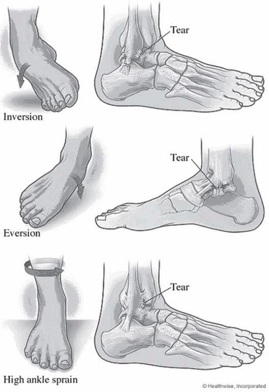 Ankle sprains are typically evidenced by pain and swelling in the ankle. A popping or snapping sensation may be felt and the person may have difficulty with bearing weight. Bruising is also common but may not be visible until hours or days after injury. The good news about ankle sprains is that most resolve with rest and support, however, what differentiates the ankle sprain that can be “walked off” almost immediately from the sprain which requires immobilization or surgical intervention? What type of support and treatment result in faster returns to activity or work?
The ankle is a complex joint where the tibia (shin) and fibula (small bone of the outer leg) meet the talus (upper-most bone of the foot.) This joint is stabilized by three main ligament complexes. The most commonly injured are those of the outside or lateral ankle. These include the anterior talofibular ligament (ATFL), calcaneofibular ligament (CFL), and posterior talofibular. These are typically injured when someone lands or rolls on the outside of their foot causing what is known as a forced inversion injury.
The inner ankle is stabilized by a number of ligaments known collectively as the deltoid ligaments. These are more commonly injured when a person rolls or land on the inner foot causing a forced eversion injury. This type of injury is also more likely to cause a fracture. The third set of ligaments is between the two bones of the lower leg and is known collectively as the syndesmosis. A high ankle sprain refers to injury to this syndesmosis.
Ankle sprains are typically diagnosed by a good history and physical examination. The physician will ask about the mechanism of injury and symptoms from the time of injury. They will examine the ankle to see where it is most tender and stress the ligaments to assess for any instability. They should also check the function of nerves and blood vessels that travel to the foot for any possible injury. X-rays may be necessary to rule out a fracture.
The Ottawa Foot and Ankle rules are a commonly used guideline for obtaining x-rays in acute injury. These guidelines state that x-rays should be obtained if the patient has tenderness over certain bone structures (medial malleolus, fifth metatarsal, or novicular), or if the patient is unable to bear weight immediately after injury or at time of examination. These are tailored to use in the emergency department and other guidelines may be used by an orthopedist or primary care provider. In those with persistent pain, an MRI to further evaluate the soft tissues may be needed.
Ankle sprains range in severity much like any other injury. Some are minor enough that even hours after injury the patient feels better. Others may last for weeks or require surgical intervention. The degree of pain and dysfunction is generally attributed to the amount of damage to the ligament or ligaments. Different grading systems can describe these in different manners.
No matter the severity of the sprain, the first measures of treatment are typically conservative. RICE (rest, ice, compression, and elevation), therapy can be helpful initially to help with pain relief and control of acute swelling. Anti-inflammatories such as ibuprophen or naproxen have also been traditionally used for pain and inflammation control. This has become more controversial in recent years as newer studies suggest that NSAID use may delay healing as inflammation is the body’s natural response to an injury in that it causes chemical signals in the body to recruit nutrients and growth factors to the site of injury. It is theorized that blocking this initial inflammation may lead to delayed healing.
Another traditional form of treatment for ankle sprains has been splinting or rigid bracing for immobilization. More recently there has been a rise in functional treatment for Grade I and Grade II sprains. Functional treatment typically consists of support with a lace-up or semi-rigid brace coupled with early physical therapy exercises to restore motion and strength. Ivins, et. al, showed people returning to sports 4.6 days sooner and work 7. 1 day sooner with functional treatment. Another study (Kerkhoff’s et. al, 2009), shows that more patients return to sport 4.88 days faster, and return to work 8.23 days faster. In general, they found that more people returned to sport in the long run.
Ankle therapy includes general range of motion as well as strengthening about the ankle. This is often combined with something referred to as proprioceptive training. Proprioception is the ability for a person to sense where they are in space. This is what allows us to catch objects in the air or step up and sown meeting the floor without falling. The nerve fibers that give us this sense are often impaired due to the ankle sprain often requiring retraining to prevent further injury from new falls or awkward positions.
Common ankle exercises for both treatment and prevention of an ankle sprain include range of motion exercises, which can simply be performed using a cloth or towel wrapped around the foot with the ends firmly in both hands. Gently pull the towel toward the face and hold for 15-30 seconds. This can be varied by also pulling with an inward roll of the ankle and outward roll of the ankle. These can be repeated 3-5 times. Also, you can perform a standing calf stretch by bending the knees while standing with their heels on the floor. Another creative way to work on ankle range of motion is to spell the alphabet with the great toe by moving the ankle through different planes.
Once you have full range of motion, strengthening exercises can be started to help prevent re-injury. Simple exercises can include pushing downward, upward, inward and outward on an immovable object holding each for 5 second for 10 sets. Then dynamic (moving), strengthening using the other foot, rubber tubing or weights can include the same motions, but instead of pushing and holding, this includes fully contracting the muscles over 1 second followed by taking 4 seconds to relax the ankle for three sets of 10 repetitions. Using the toes to pick up small objects and walking forward and backward on toes and heels are also common strengthening exercises.
Proprioceptive training is typically started when the patient can weight bear without much pain. This can be as simple as heel raises and toe raises or as involved as using a wobble board as well as walking on different surfaces (Berkowitz & Fierstein, 2011). Proprioception is also improved with single leg balance and using a jump rope. Formal physical therapy is often needed to initiate different exercises although most can be done at home. A good strengthening program helps facilitate your return to the court.
Achilles Injuries
October 5, 2015
Preventing Achilles Injuries: What can go wrong on the court?
Most people are familiar with the great story of Achilles who was dipped into the River Styx to become invincible. Unfortunately, Thetis held him at the ankle and so is born the “Achilles heel.” According to Homer’s Illiad, Achilles was slain when Paris shot an arrow into his heel. Fortunately, flying arrows are rare in today’s world, but that does not mean that you are immune to Achilles injuries on the court.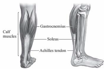
The Achilles tendon inserts on the back of the heel (or the calcaneus). It is comprised of two muscles (the gactrocnemeus and the soleus) which merge to become one tendon. It becomes round and then flattens about 4 cm. proximal to the heel. There are essentially four Achilles injuries that can arise with the tendon: Achilles tendinitis and tendinosis, paratenonitis, insertional tendinitis and Achilles ruptures.
Achilles Tendonitis
Achilles tendonitis is a condition when the Achilles is irritated and inflamed. There is a relatively avascular zone in the Achilles tendon that is roughly 2-6 cm. for the insertion of the Achilles into the heel. It is a common injury in recreational athletes and occurs when there is overuse of the tendon that causes inflammation leading to pain and swelling. When the condition is chronic, it leads to Achilles 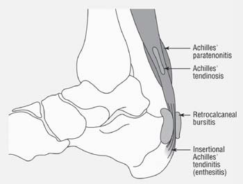 tendinosis. With tendinosis, there are microscopic tears within the tendon. The paratenon is a sheath that envelopes the tendon. It can become inflamed with repetitive strain or overuse. The tendon may also be thickened. The pain improves with rest and is aggravated with activity. It tends to be associated with overuse and is not a precursor to Achilles ruptures. Insertional tendinitis is characterized by inflammation and pain surrounding the point where the tendon inserts into the heel. This can lead to partial tearing or rupturing of the tendon. A person will complain of tenderness directly over the Achilles insertion into the heel and can make weightbearing difficult. The tendon can become hardened and thickened. Surgery may be necessary for those cases recalcitrant to nonoperative treatment such as rest, cross-training, anti-inflammatories, stretching, and physical therapy.
Achilles Tendon Ruptures
Achilles tendon ruptures can occur in the substance of the tendon (in the avascular zone) or they can occur where the tendon avulses off the calcaneus. They are up to 12 times more common in men than women and usually result from noncontact injuries where the load to the Achilles exceeds the strength of the tendon. This can result from mechanisms such as a forceful push-off of the foot with an extended knee (jumping or sprinting), sudden unexpected ankle dorsiflexion, or violent dorsiflexion of a plantar-flexed foot.
Many times, a person may feel like they got kicked in the back of the heel by someone. The immediate pain is shortlived, but people will notice the inability to push the heel off the ground when walking. There may be swelling and/or bruising. Sometimes, a person may notice a palpable defect in the tendon. It is usually difficult to walk. When a physician squeezes the calf in the normal ankle, the foot will dorsiflex or flex down toward the ground. When the same test is done in the injured leg, there is no movement at the ankle. This is called a positive Thompson test. Achilles ruptures are almost always diagnosed clinically, but when there is uncertainty, an MRI is an excellent test to evaluate the integrity of the Achilles tendon.
Despite recent attention to surgical treatment of Achilles ruptures, the decision to treat these injuries surgically versus nonoperatively remains controversial. Nonoperative management is historically associated with a high risk of Achilles rerupture, ranging from 13 to 30%, but without wound complications. Operative management of Achilles tendon ruptures results in a lower re-rupture rate (0 to 6%), earlier mobilization, and better return to sports, but with the risk of higher wound complications (up to 21%). Recent studies suggests an infection rate that is much lower.
The goal of surgical management is early mobilization to maximizes healing potential. This requires strong suture material and a sound technique. Two such techniques include the Krackow suture and the “Giftbox” technique. as described by
Labib, et al.
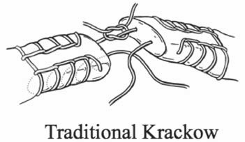
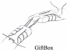
After surgery, the repair is splinted for up to two weeks with no weight bearing. Thereafter, the heel is placed in a boot with heel lifts that are removed over the next month. Weight bearing usually begins around two weeks, although this may vary depending on the surgeon.
Achilles injuries occur as a result of being in the right situation at the wrong time. That is, the force applied to the tendon is stronger than the tendon can withstand. Sometimes, that just cannot be prevented. Achilles tendon injuries such as tendinitis, insertional tendinitis, tendidnosis, and paratenonitis typically respond well to nonoperative management.
Prevention of Achilles Injuries:
As people age, calf and Achilles tightness is almost inevitable due to several reasons. First, as we become more sedentary, we have less daily stretch of the calf muscles and Achilles tendons. There is also age-related decrease in the elasticity of the Achilles tendon. Finally, higher-heeled shoes put the Achilles tendon in a shortened position. A good stretching and strengthening program is the best way to prevent Achilles injuries.
Achilles tendon stretching will result in satisfactory relief in better than 90% of the people suffering from Achilles pain. Some
people require as little as 3 weeks to see improvement while others may need upwards of 5-6 months to break through a long standing Achilles contracture. The heel stretching protocol is outlined below in detail. In the beginning, the amount you stretch will vary due to pain or soreness of the Achilles. Be sure to stretch both feet.
First of all, you will need a step to stand on with a wall for support. A routine step lift works extremely well. With your back against the wall and your knees straight, place the balls of your feet on the step and slowly relax your ankles, letting your heels go downward (see figure). You should not be gripping the step with your toes. If you are in the correct position, you should feel a pulling or tightness in your upper calf muscle, just below the knee. This should be tolerated until slight pain is felt.
For best results, do the stretch three times per day, gradually increasing the amount of time. You may need to stay at the same amount of hang time for a few days. Increase your time gradually, maxing out at three minutes. This is a gradual process and be patient. If you go right to three minutes, you may cause yourself more pain. Be Patient!
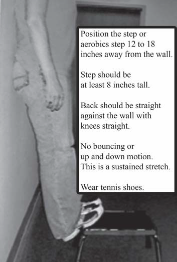 Week I – 15 seconds, 3 times/day
Week II – 30 seconds, 3 times/day
Week III – 1 minute, 3 times/day
Week IV – 1 1/2 minutes, 3 times/day
Week V – 2 minutes, 3 times/day
Week VI – 2 1/2 minutes, 3 times/day
Week VII – 3 minutes, 3 times/day
Hand Only CPR: A Better Way to Save Lives?
October 5, 2015
Hand Only CPR: A Better Way to Save Lives?
Out-of-hospital cardiac arrest affects more than 300,000 people annually in the United States. It is certainly something that could happen on the handball court. When this happens, the heart stops beating. A person usually collapses, stops breathing and is unresponsive. Since survival is so strongly dependent on immediate action by bystanders, what if we could increase survival odds through hand only CPR?
Although conventional CPR (chest compression and rescue breaths) can double the survival rates of victims having cardiac arrest, only one third of the victims have someone attempt CPR. The most common barrier to bystander action is the their concern for disease transmission as a result of mouth-to-mouth resuscitation. Another barrier is the fear of harming the victim from inadequate training. Much attention has been given to eliminating the barriers and getting more people to help begin resuscitation.
Hand only CPR (chest compressions only) has increased bystander involvement as well as survival rate. Several studies have shown that hand only CPR can be as effective as conventional CPR in the out-of-hospital setting. As a result, The American Heart Association released an advisory statement supporting hand only CPR in an attempt to increase bystander response to victims suffering from cardiac arrest. In fact, people were more likely to perform hand only CPR over conventional CPR regardless of their basic life support training. A study out of Arizona supports hand only CPR over conventional CPR. After looking at 4,415 adult cardiac arrests outside of hospitals from 2005 to 2009, researchers found that the rate of bystanders attempting CPR increased from 28% in 2005 to 40% in 2009. People were more likely to use hand only CPR.
In addition, victims were more likely to survive. Thirteen percent (113/849) of victims who received hand only CPR survived compared to eight percent (52/ 666) who received conventional CPR. In a recent study in The Lancet, three studies were combined and showed a survival rate of 14% for hands-only CPR versus 12% for conventional CPR. The authors recommended that rescuers should focus on hand only CPR with out of hospital cardiac arrest. So how do you do hand only CPR? First, if someone collapses, call 911 or have someone else call for you. Then get directly over the victim, lock your elbows and push hard in the center of the chest with both hands at a rate of approximately 100 times per minute. If you know standard CPR, then include rescue breaths. If an AED is available, switch it on and follow the instructions. Remember, any attempt at CPR is better than no attempt. According to the American Heart Association, hand-only CPR is recommended for use in adults who suddenly collapse. Conventional CPR is still recommended for “all infants and children, adults already found down or not breathing normally, and any victims of drowning or collapse due to breathing problems.”
Automatic Electronic Defibrillator (AED) Cardiac arrest usually results from an abnormal heart rate called ventricular fibrillation. It can happen to anyone at anytime. A automatic electronic defibrillator (AED), gives a victim an electronic shock that corrects the ventricular fibrillation by interrupting the irregular rhythm of the heart, reestablishing the normal electrical rhythm. An AED is a device that allows a person to monitor the heart rhythm of a victim and delivers a shock if necessary to re-establish the normal rhythm. Using AEDs with CPR quadruples the survival rate compared with using CPR alone. AEDs are easy to use. and all of them come with instructions that should be used. Place one pad on the upper right chest and one pad on the lower left side. If the electrodes are not attached properly, there is usually an error message. When the AED is attached properly, it will prompt you to analyze the rhythm. If the heart rhythm requires defibrillation, you will be prompted to do so. Be certain nobody is in contact with the victim while administering the shock. A simple way to make sure that every is safe is to say “I’m clear, you’re clear, everybody clear!” There are some special considerations with AEDs. If the victim is hypothermic, it may take longer to assess for a pulse (up to 45 seconds). If patients have pacemakers or implantable devices, do not place the AED pads over the device. Some patients use nitroglycerin patches and these should be removed prior to applying the AED pads.
Shoulder Treatment Education
October 5, 2015
Why an Article on Shoulder Treatment Education?
Proper shoulder treatment education is important when you or someone you love has a shoulder injury. The shoulder is a very delicate joint. Simple tasks like picking up pans, opening a window, moving a chair, or carrying a child can cause shoulder injuries. Activities like tennis, football, volleyball, and martial arts can cause further shoulder damage. Unfortunately, simply aging can cause tendon pain in the shoulder. Below are some of the common treatment options. Shoulder treatment education would not be complete without mentioning some of our non-surgical options, but we will get into those in a later post.
What is Shoulder Arthritis?
Arthritis is a common disease that affects the shoulder, causing joint pain, stiffness and swelling. When a person gets arthritis, they lose cartilage or the cushion in the joint. Cartilage allows the joint to glide easily during motion. When cartilage wears, there is bone on bone rubbing which can be painful. In addition, the joint becomes inflamed and there is usually a restriction of motion.
How is Shoulder Arthritis diagnosed?
Many patients will complain of a deep ache that can radiate to the lateral arm. The pain usually gets worse with movement or activity. With time, there may even be pain at rest and eventually, patients will awaken at night with shoulder pain. Patients will complain of grinding and difficulty with motion. In the office, a physician will diagnose arthritis through a thorough physical exam and taking the proper x-rays.
What is Shoulder Replacement Surgery?
In total shoulder replacement, the damaged surfaces of the humeral head and glenoid socket are resurfaced with metal and plastic implants. The humeral head is replaced with a metal component and the glenoid socket is replaced with a polyethylene component that is glued in with bone cement. When both sides of the shoulder are replaced, it is called a total shoulder replacement. When only the humeral head is replaced, it is called a shoulder hemiarthroplasty or a partial shoulder replacement.
Why should I consider Shoulder Replacement Surgery?
Shoulder replacement surgery should be performed to alleviate pain and improve function. It is an excellent option for patients that have failed other conservative measures such as physical therapy and antiinflammatory medication.
How long will my Shoulder Replacement last?
Anytime implants are used to replace your normal anatomy, it is expected that they will wear with time. The survivorship of a shoulder replacement is up to 93% at 10 years and 87% at 15 years. (Torcia et al.) At one year from the time of surgery, 98.5% patients are glad they had the procedure done. Ninety percent of patients feel that their new shoulder has attained the 80 percent of what they feel a “normal” shoulder would be and 37 percent of patients think that their new shoulder is normal. (Warner et al.)
What are my restrictions after a Total Shoulder Replacement?
Most activities can be resumed after a shoulder replacement. A person usually resumes activities such as swimming, golf and tennis by six months after surgery. Activities that involve major impact (such as contact sports or those where falls are frequent) or heavy loads (such as lifting heavy weights) should be avoided since these may increase the chance of rotator cuff tears, hardware loosening, increased wear and/or fracture.
What will be my recovery time?
Patients whom undergo total shoulder replacement usually stay in the hospital for 1-3 days after surgery. Physical therapists will begin moving the shoulder on the first postoperative day and patients will wear a sling for the first 4-6 weeks. At that time, patients can use their shoulder as tolerated with a weight restriction of 5 pounds. Most activities can resume in 4-6 months.
What are the risks of Shoulder Surgery?
Complications are rare, but they are also real and do happen. Some of the more common complications include infection, stiffness, instability, component failure, fractures around the prosthesis and failure to get complete pain relief. Fortunately, the benefits of shoulder replacement far outweigh the risks.
What is the difference between a chronic torn rotator cuff and acute tear?
Do both require surgery?
An acute rotator cuff tear is a tear that occurs after an injury. There is usually sudden pain in the shoulder accompanied by limited movement and weakness. A chronic tear is a tear that is greater than 6 to 12 weeks old. They usually involve the dominant arm and gradually worsen. People may develop weakness. There are many functioning patients with chronic tears. At sixty, fifty percent of asymptomatic patients would have a partial thickness rotator cuff tear if they had an MRI. At 80, fifty percent of patients would have a full thickness tear (all the way through). Chronic tears usually require surgery if the patient has persistent pain or weakness despite 6-12 weeks of physical therapy. Most acute rotator cuff tears or tears in young patients should be fixed surgically.
Why do physicians recommend a total shoulder replacement now?
Ten years ago, I never heard of a total shoulder. Shoulder arthritis is a common disease affecting thousands Americans annually. Many people with shoulder arthritis will have adequate pain control and function using anti-inflammatory medication and physical therapy. For patients that fail to improve with non-operative management, shoulder replacement is an excellent alternative. Up to 40,000 shoulder replacements are performed annually. At one year from surgery, 98.5% patients state that they are glad they had their shoulder replaced and would do it again. The successful results of shoulder replacement are similar to those for hip replacement surgery.
Do you have to repair a torn bicep even if it was injured five months ago?
The biceps is a muscle on the front of the shoulder that allows a person to flex and supinate the elbow. It has two muscle bellies, the long head of the biceps that inserts on the labrum at the top of the shoulder socket and the short head of the biceps that inserts on the front of the scapula at its coracoid. At the elbow, the bicep muscle bellies merge into one tendon that inserts into the radius. When a patient ruptures the distal part of the biceps, they may lose up to 40% of their strength with flexion and supination. Surgery is typically recommended when it is the dominant arm and the patient’s job requires significant use of the arm. Surgery is not always recommended for ruptures that occur near the shoulder at the long head of the biceps. Most people respond well to physical therapy.
I am a senior and I don’t want surgery but I can’t lift my arm, would therapy help relieve the pain?
There are many reasons a person may have pain in the shoulder and not all of them need to be treated surgically. Most shoulder ailments improve with physical therapy. It is helpful to have a thorough physical exam so that physical therapy can be guided appropriately. Sometimes, a platelet injections (platelet injections) or steroid injection can be helpful.
We hope this shoulder treatment education article has been helpful! For any additional questions or treatment options, give us a call at (513) 354-3700 or schedule an appointment with Dr. Robert Rolf!
Orthopaedic Research & Articles
October 5, 2015
Beacon Orthopaedics and Sports Medicine Encourages Orthopaedic Research & Articles:
Continuous research and education is a key part for all of the doctors at Beacon. This is very evident in the work and contributions of Dr. Adam Miller. Through research and engineering, improvements in technology, new treatment options are developed for various conditions. By contributing to Orthopaedic Research & Articles, Dr. Miller is able to offer innovative and advanced care.
Below are several links to articles or research to which Dr. Miller has collaborated or contributed. Select any of the links in this section to learn more about Dr. Miller’s academic research.
- Delasotta LA, Orozco F, Miller AG, Post Z, Ong A. Distal femoral fracture during primary total knee arthroplasty. J Orthop Surg (Hong Kong). 2015;23(2).
- Raikin SM, Miller AG, Daniel J. Recurrence of Hallux Valgus: A Review. Foot Ankle Clin. 2014.
- Orozco F, Post ZD, Baxi O, Miller A, Ong A. Fibrosis in hepatitis C patients predicts complications after elective total joint arthroplasty. J Arthroplasty. 2014;29(1):7-10.
- Miller AG, Schon LC. Fixation of Midfoot and Forefoot Fractures. Techniques in Orthopaedics. 2014;29(1):24-32.
- Miller A, Raikin SM. Lateral Ankle Instability. Operative Techniques in Sports Medicine. 2014;22(4):282-9.
- Miller AG, Raikin SM, Ahmad J. Near-Anatomic Allograft Tenodesis of Chronic Lateral Ankle Instability. Foot & ankle international / American Orthopaedic Foot and Ankle Society [and] Swiss Foot and Ankle Society. 2013 Jun 14. Epub 2013/06/19.
- Miller AG, McKenzie J, Greenky M, Shaw E, Gandhi K, Hozack WJ, et al. Spinal Anesthesia: Should Everyone Receive a Urinary Catheter? The Journal of Bone & Joint Surgery. 2013;95(16):1498-503.
- Miller AG, Daniel JN. Achilles tendon pathology associated with tophaceous gout infiltration. Current Orthopaedic Practice. 2013;24(1):103-4.
- Garras DN, Durinka JB, Bercik M, Miller AG, Raikin SM. Conversion Arthrodesis for Failed First Metatarsophalangeal Joint Hemiarthroplasty. Foot & ankle international / American Orthopaedic Foot and Ankle Society [and] Swiss Foot and Ankle Society. 2013 Apr 23. Epub 2013/04/25.
- Miller AG, Slenker N, Dodson CC. Terrible triad of the shoulder in a competitive athlete. Am J Orthop (Belle Mead NJ). 2012 May;41(5):228-9. Epub 2012/06/21.
- Miller AG, Purtill JJ. Total knee arthroplasty component templating: a predictive model. J Arthroplasty. 2012 Oct;27(9):1707-9. Epub 2012/05/29.
- Miller AG, Purtill JJ. Accuracy of digital templating in total knee arthroplasty. Am J Orthop (Belle Mead NJ). 2012 Nov;41(11):510-2. Epub 2013/02/23.
- Miller AG, Margules A, Raikin SM. Risk factors for wound complications after ankle fracture surgery. J Bone Joint Surg Am. 2012 Nov 21;94(22):2047-52. Epub 2012/11/23.
- Miller AG, Bercik MJ, Ong A. Nonagenarian hip fracture: treatment and complications. J Trauma Acute Care Surg. 2012 May;72(5):1411-5. Epub 2012/06/08.
- Garras DN, Hansen PL, Miller AG, Raikin SM. Outcome of modified Kidner procedure with subtalar arthroereisis for painful accessory navicular associated with planovalgus deformity. Foot Ankle Int. 2012 Nov;33(11):934-9. Epub 2012/11/08.
- Bercik MJ, Miller AG, Muffly M, Parvizi J, Orozco F, Ong A. Conversion total hip arthroplasty: a reason not to use cephalomedullary nails. J Arthroplasty. 2012 Sep;27(8 Suppl):117-21. Epub 2012/05/29.
- Aynardi M, Miller AG, Orozco F, Ong A. Effect of work-hour restrictions and resident turnover in orthopedic trauma. Orthopedics. 2012 Nov;35(11):e1649-54. Epub 2012/11/07.
- >Parvizi J, Miller AG, Gandhi K. Multimodal pain management after total joint arthroplasty. J Bone Joint Surg Am. 2011 Jun 1;93(11):1075-84. Epub 2011/06/10.
- Miller AG. Weighing your future job options in today’s market. Am J Orthop (Belle Mead NJ). 2011 Nov;40(11):E241-2. Epub 2012/01/21.
- Miller AG, Swank ML. Dermabond efficacy in total joint arthroplasty wounds. Am J Orthop (Belle Mead NJ). 2010 Oct;39(10):476-8. Epub 2011/02/04.
- Swank ML, Miller AG, Korbee LL. Transition Zone Failure in Patients Undergoing Instrumented Lumbar Fusions from L1 or L2 to the Sacrum. 2007.
Surgical Videos (Graphic)
October 2, 2015
At Beacon Orthopaedics & Sports Medicine, we provide an array of graphic surgical videos to educate patients and medical professionals about various orthopedic procedures. These videos offer a transparent look into the surgical process, aiding in informed decision-making.
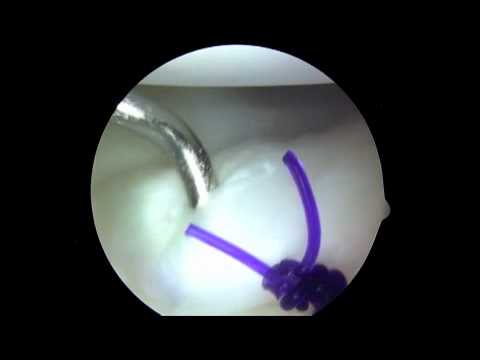
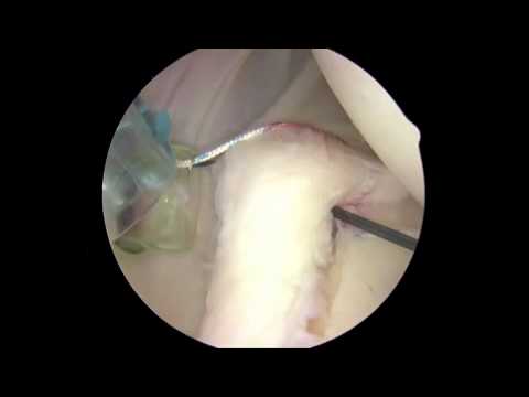
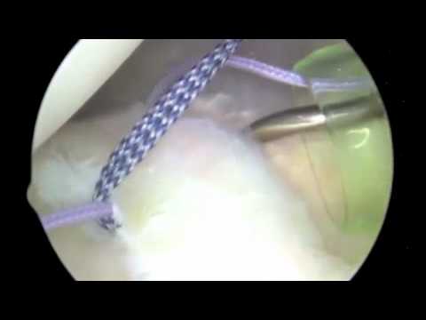
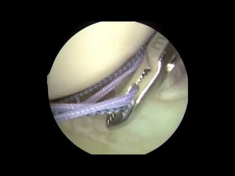
https://www.youtube.com/watch?v=6kp2RLjVQ2c
https://www.youtube.com/watch?v=MevcmzIPNM0
Pre-Op & Post-Op Instructions
October 2, 2015
What You Need To Know About Knee Arthroscopy:
On the day of surgery you cannot eat or drink anything after midnight the night prior to your procedure. Only take medications that the preadmission testing nurse has told you were ok to take with a sip of water. Please shower the morning of your surgery but do not apply any lotions or perfumes after your shower. It’s a good idea to leave any jewelry at home since you will be asked to remove it prior to going into the operating room. The anesthesiologist will meet you prior to your  surgery and will keep you comfortable. Dr. McClung will see you and mark the correct limb prior to surgery.
During your surgery you will be completely asleep, under a general anesthesia. The entire procedure will last between 30 and 45 minutes. With arthroscopy, we can fix problems inside the joint with just making 2 or 3 small incisions. A small camera is inserted into the joint through one of these holes so we can look at the bones and tissues in the joint. Small tools are then inserted through the other hole. During your surgery, it is possible that Dr. McClung will trim or repair a torn meniscus, smooth any rough surfaces, clean up any arthritis or loose pieces of bone and replace your ACL or PCL if you’ve discussed this with him prior to surgery. You will go home the day of surgery, but will need a responsible adult to take you home since you will still have anesthetic in your system. You will need 24 to 48 hours after surgery to get the anesthesia out of your system, it’s a good idea to plan to have this time off work. If you have a desk job, it’s acceptable to return to work within 3-5 days after surgery; if you have a more strenuous job, you should discuss this with Dr. McClung prior to scheduling your surgery.
Patients can click here to download and/or print pre-operative instructions for knee arthroscopy.
Knee arthroscopy Post Operative Instructions from Dr. Glen McClung:
- Sponge bathe until your dressing change appointment 2 days after surgery. Once your dressing has been changed you may shower as long as your incision sites are covered with waterproof bandaids. Remove and replace your waterproof bandaids daily after bathing.
- Do not put any ointment or peroxide on the incision sites. Normal drainage appears bloody or like water, if there is any abnormal drainage from the incision site please let us know.
- Do not submerge in a bathtub, hot tub or pool for 4 weeks post operatively
- Wear your sling or brace at all times as it was put on in the operating room unless you were instructed otherwise by Dr. McClung.
- Only take medication as prescribed by Dr. McClung. Do not take it more often than prescribed. If your pain is not controlled please call our office, do not take any additional Tylenol. If you need a refill on your medication, call the office. Please understand that it may take up to 24 hours after your call for your medication to be refilled.
- Ice the operative site for 20 minutes at a time with a traditional ice bag or for one hour with the ice machine. Do not place the ice bag or ice machine directly on the skin, place a towel between the ice bag and your skin. Icing the operative site regularly will decrease swelling and pain to the area.
- You will go home from surgery with stockings on your legs to prevent blood clots. These may be removed once you are getting up and walking around or getting around on your crutches regularly.
Thank you for choosing Beacon Orthopaedics and Sports Medicine and Dr. McClung for the care of your orthopaedic injuries. We look forward to serving you and getting you back to a pain free lifestyle!
Patients can click here to download and/or print post-operative instructions.
Sleeping After Shoulder Surgery
September 30, 2015
Dr. McClung’s tips for sleeping after shoulder surgery
The most common objective of shoulder surgery is to relieve pain. In order to facilitate quick and correct healing, as well as to avoid future pain, here are some suggestions for sleeping after shoulder surgery. Improper posture or poor positioning can increase recovery time or cause damage to the affected area.
- I suggest sleeping in a reclined position. This can be achieved by sleeping in a recliner for the first couple weeks after surgery.
- Avoid sleeping flat on your back. Bolstering yourself up in your bed with several pillows or with a 45 degree wedge that you can purchase at a medical supply store will help you rest. Often, patients have to sleep with an incline for 4 to 6 weeks after surgery.
- Wear your sling. Wearing your sling while you sleep keeps tension off the repair that was done in your shoulder. It also helps patients to avoid putting their shoulders in painful positions while they are sleeping.
- Purchase an Iceman. Though it is a large out of pocket purchase, using a cryocuff or “iceman” will significantly decrease your pain. You can use this as often as you would like for as long as you would like. The water must be changed every 40-60 minutes but, many patients find it’s easier to fall asleep with the cuff on their shoulder.
- Time your medications. Planning our when you take your medications can be an important part of sleeping after shoulder surgery. This is especially true if you find yourself in a lot of pain. Time your pain medication to be taken 30 minutes prior to when you are planning to go to sleep.
Hopefully these suggestions for sleeping after shoulder surgery are useful to you. If you have any questions about recovery times or other activities after surgery, please call Beacon Orthopaedics at (513) 354-3700.