Imaging in Orthopedics: What’s the right image for you?
November 30, 2021
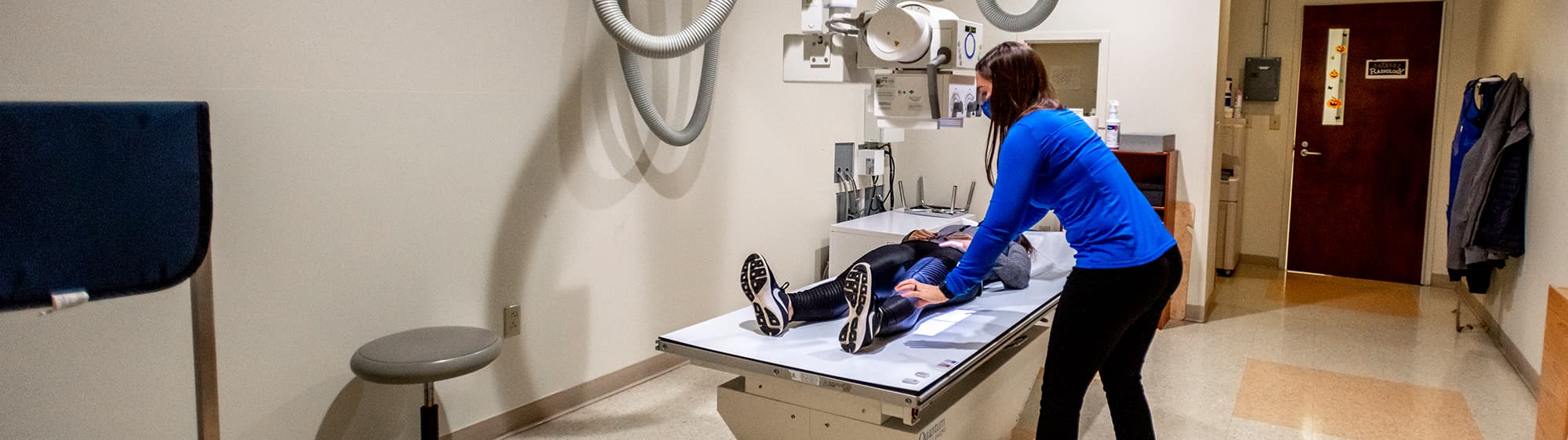
When you go to an Orthopaedic doctor for foot and ankle care it is very likely you will be getting some sort of imaging done. When evaluating a patient, aside from the patient’s history and exam, an Orthopedic surgeon uses imaging to guide treatment. In most cases, doctors can tell a lot from basic plain films or radiographs (X-Rays). These tests help the doctor in their guidance with the next step for the patient.
The vast majority of patients can be treated without advanced imaging and use radiographs to treat them appropriately and successfully. Specifically in lower extremity orthopedics, it is important to get weight bearing X-Rays. These will help show the alignment of the leg, ankle, and foot, while guiding the doctor towards the process of how to treat the patient and what they may need to correct. In some cases, advanced imaging may be recommended to help determine the nature of the problem. These include: magnetic resonance imaging (MRI), computed tomography (CT), ultrasound and bone scans. Below are many of the imaging techniques used by foot and ankle specialist, Dr Adam Miller, to help determine the next step in his patient’s journey. Here is what you can expect.
X-Ray: The most common imaging technique. X-ray uses small amounts of radiation to detect the mineral in bone. This image is captured for the orthopaedist to view. They help detect fractures and more specifically the type of fracture. Fractures can have different characteristics based on the timing. This helps Dr. Miller determine if the fracture is healing and how old the fracture is. With these findings there are certain hallmarks to look for in determining what the fracture looks like. In some more rare diagnoses such as oncology or cancer, there are specific characteristics we look out for to make sure there is nothing abnormal going on that would be of concern. X-Rays are sometimes helpful to determine metal foreign bodies.
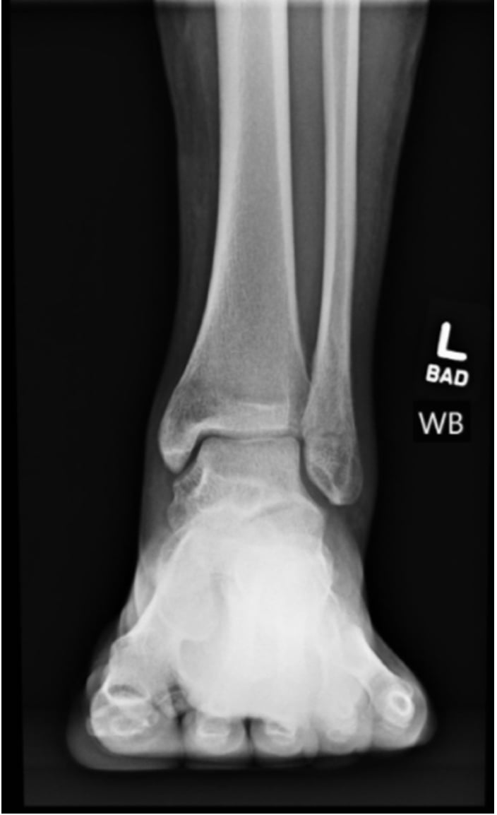
In orthopaedic foot and ankle care, X-Rays help evaluate the alignment of the patient’s bones. One can also determine the amount of chronic changes such as arthritis. These X-Rays are then used to set a plan with the patient. Follow up X-Rays subsequent visits are used to check on a surgery outcome or to check on healing of an injury. The time continuum of the X-Rays at each visit helps us know how the healing is going.
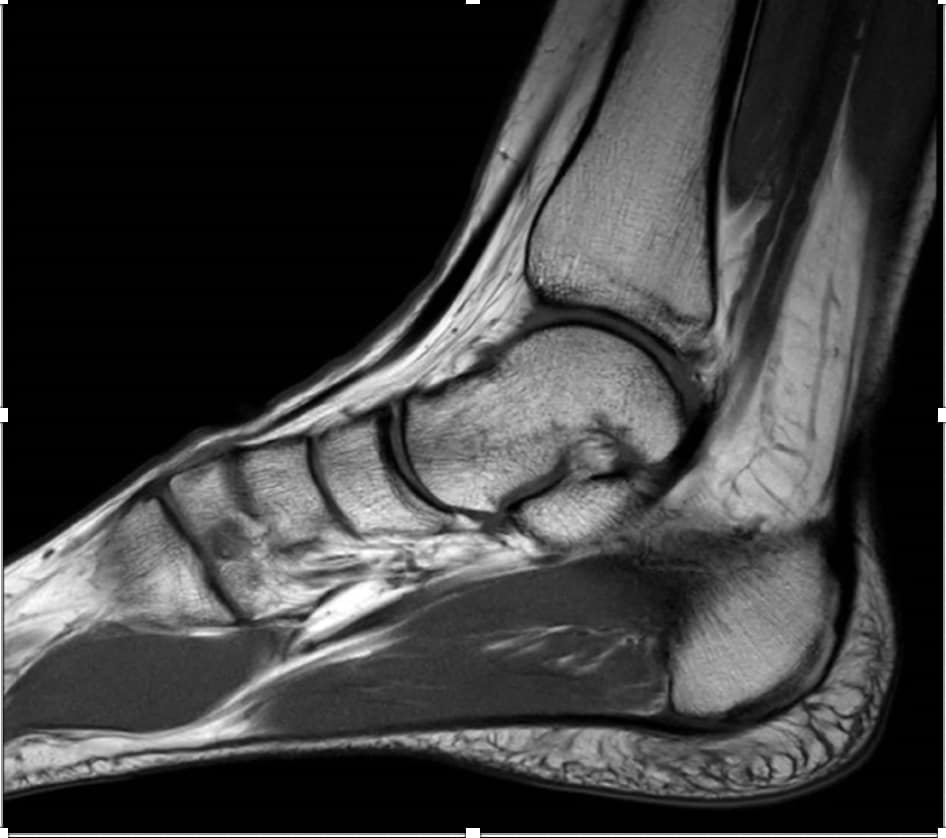
MRI: The second most common study is an MRI or Magnetic Resonance Imaging. No radiation is required. Instead, this uses magnetic fields to create detailed images on a focal part of the body. MRI’s give very detailed imaging of essentially everything: bone, soft tissue and can determine the difference between tendons, ligaments, bone, fluids, and solids. These are very helpful with subtle injuries but also soft tissue injuries that the X-Rays can’t pick up. For example, if you were to have the following: sprained ankle, cartilage injury, subtle bone injury such as a stress injury, or different changes of arthritis. All these injuries can be picked up on an MRI much more accurately. For example: an X-Ray may not be able to show a stress fracture for 2 weeks before any subtle changes occur. But on an MRI within a few days, it is over 95% accurate for a stress injury. These are often obtained to make a diagnosis or plan for a surgery.
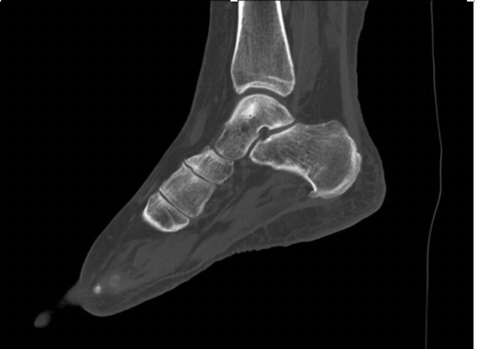
CT: A CT Scan (Computed Tomography Scan) is a 3-D scan of bone and soft tissue which uses radiation instead of magnetic imaging. Orthopaedists use CT’s when they’re trying to see very fine bone detail. Certain bone specific diagnoses require a CT to see more bone detail than what an MRI can give you. A CT is also best used in cases where a patient already has metal in their body that limits the field of view on an MRI. Sometimes we use CT to determine healing and if a certain bone healed completely.
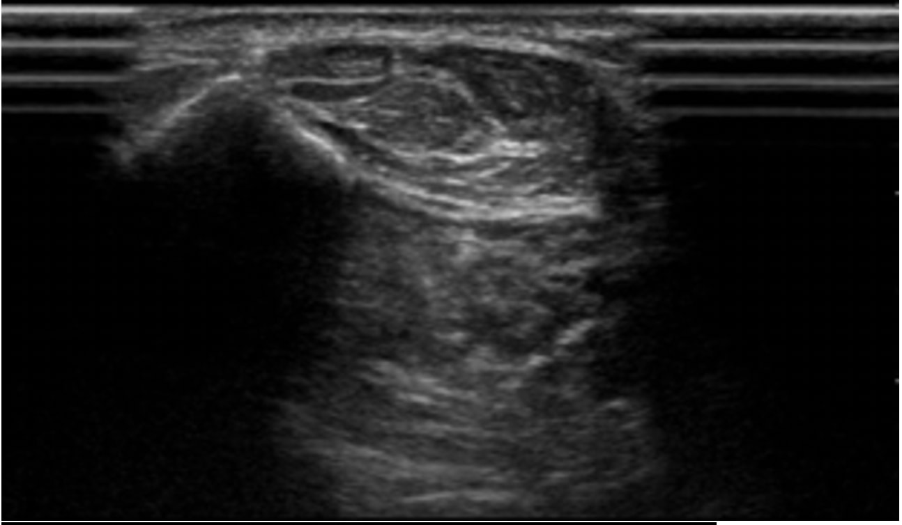
Ultrasound: An Ultrasound is a dynamic study. Unlike other imaging studies, the imaging is obtained in real time allowing the ultrasonographer to have the patient move and react to what they see. This allows us to check if anatomy is moving correctly and how structures look in different orientations. Usually ultrasound is performed when the patient is active in the office, and is used to look at anatomy while the patient is moving. It also allows us to look at different areas on the patient at one time. This can help to really see the movement of the anatomy such as a tendon or bones coming together. It is helpful to perform injections in certain joints that are difficult to identify skin deep. We use these for different steroid injections and other types of injections.
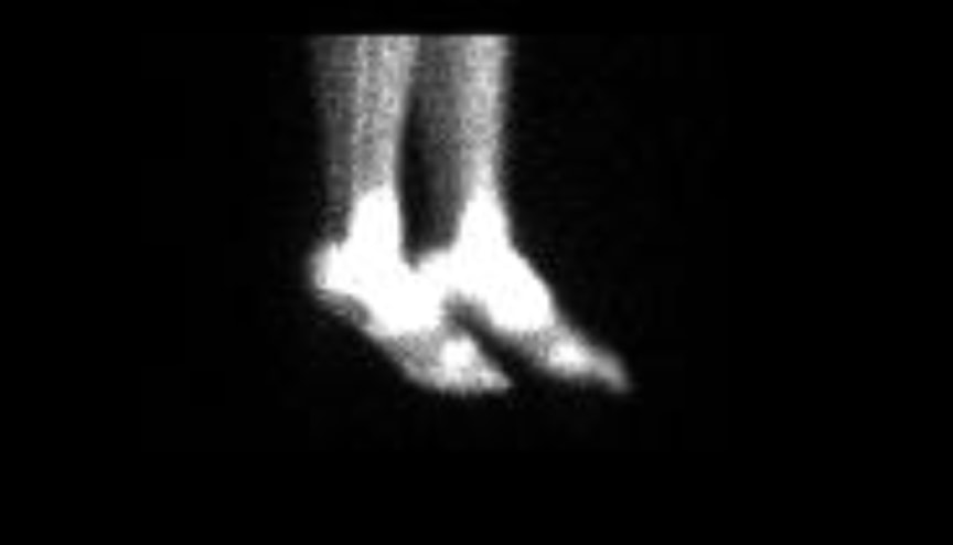
Bone scans are a group of studies using an injection in the body through a vein. This fluid spreads throughout the body and pools in active areas of the bone. How the fluid acts gives clues to various diagnoses. There are several uses in orthopaedics such as periostitis, stress fractures, inflammation, infection, and cancer.
If you think you may need imaging done do not wait, we are here to help! Dr. Miller is Beacon Orthopedics foot and ankle specialist and is available at several locations around the Cincinnati area. Contact us today for more information! Click here to learn more and schedule an appointment with Dr. Miller for your foot and ankle injuries.